Approach
Diagnosis of the correct gestational age involves a detailed review of the maternal medical history, including any available antenatal records, physical examinations, and fetal ultrasound, prior to delivery. This review is followed by clinical assessment of the newborn gestational age.
Resuscitation should be provided as necessary in line with local guideline recommendations.[28][29]
Early consultation with a neonatologist is recommended, as specialised care for premature neonates is required in most cases.
Antenatal history
A detailed maternal medical and obstetric history should be obtained and reviewed, including:
Antenatal care visits
Last menstrual period (LMP)
Blood type
Age
Parity
Current medications
Antenatal serology and cultures (e.g., HIV, syphilis, hepatitis B, and hepatitis C status)
Evidence of colonisation with group B Streptococcus
Any fetal abnormalities identified on antenatal screening
Duration of ruptured membranes
Presence of intrapartum meconium
Maternal anaesthesia
Evidence of fetal distress.
Early (6 to 12 weeks' gestation) ultrasound is the most accurate means of gestational age determination antenatally.
In many cases, due to limited resources, gestational age is estimated, often less accurately, from the last menstrual period (LMP) or the estimated fetal weight just prior to delivery.[30] Gestational age is estimated as the number of weeks since the LMP ± 2 weeks. LMP gestational age assessment may be incorrect by as much as 2 weeks due to variations in individual menstrual cycle length, recent hormonal contraception, first-trimester bleeding, and inaccurate maternal recollection of dates.
Examination of the mother prior to delivery
The distance in centimetres from the maternal superior pubis to the uterine fundus is roughly equivalent to the fetal gestational age. However, several factors, such as uterine fibroids or malposition, maternal body habitus, multiple gestation, oligohydramnios or polyhydramnios, fetal growth restriction, or the presence of a small-for-gestational-age infant, may result in an inaccurate assessment of gestational age using this method.
Examination of the newborn
Gestational age determination is important for designating further care of the premature newborn and is necessary for appropriate counselling of parents. There is wide variation by gestational age in the severity of outcomes such as overall survival, immediate post-delivery medical problems, treatment and necessary procedures, length of hospital stay, and complications of prematurity.
Extreme prematurity: gestational age below 28 weeks
Birth weight is typically <1000 g.
Infants have decreased tone and extended rather than flexed extremity posture with a wide range of motion throughout.
They have little lanugo hair coverage over most of their bodies, no vernix caseosa, thin transparent/pink skin with visible veins, limited or no plantar creases, flat areolae with nearly imperceptible breast tissue, fused or open eyelids, folded ear pinna cartilage, preterm genitalia (male: scrotum empty with faint or no rugae, female: prominent clitoris and small or flat labia minora).
Severe prematurity: gestational age 29 to 31 weeks
Birth weight is typically 1000 to 1500 g.
Infants exhibit limited flexor posture with moderate range of motion throughout.
They are generally covered in lanugo with variable amount of vernix, have smooth pink skin with many visible veins, no plantar creases or faint markings, no areolar buds, open eyelids, folded or limited-recoil ear pinna cartilage, preterm genitalia (male: testes in upper canal with minimal scrotal rugae, female: prominent clitoris with small/enlarging labia minora).
Moderate prematurity: 32 to 33 weeks
Birth weight is typically 1500 to 2000 g.
Infants have mild flexor tone and mild range of motion throughout.
They may have abundant or thinning lanugo with variable amount of vernix, few visible veins and some superficial skin peeling, faint red marks or an anterior transverse crease on plantar surface, rarely present areolar buds, open eyes, ears with some elastic recoil to pinna, preterm genitalia (male: testes palpable in canal or high in scrotum with rare to few rugae present; female: prominence of clitoris is waning and labia may be of equal size).
Late-preterm: 34 to 36 weeks
Birth weight is typically 2000 to 2750 g.
Infants appear smaller but very similar to those born at term, an appearance that puts them at risk for complications as they are not yet fully mature. They exhibit moderate flexor tone in upper and lower extremities with limited range of motion.
They have superficial skin peeling to cracking with rare veins, thinning lanugo with bald areas, few creases on anterior plantar surface, small areolar buds, open eyes, ear pinna with ready elastic recoil, more mature genitalia (male: descended testes, with scrotal rugae present; female: equally prominent labia majora and minora).
Term: 37 to 42 weeks
These infants are in flexor posture and have limited range of motion.
They have variable degrees of skin cracking or peeling with no visible vessels, plantar creases cover the majority of the sole, full areolae with prominent buds, open eyes, formed pinna with immediate recoil, term genitalia (male: testes descended into the scrotum with prominent rugae; female: large labia majora and small labia minora).
New Ballard Score
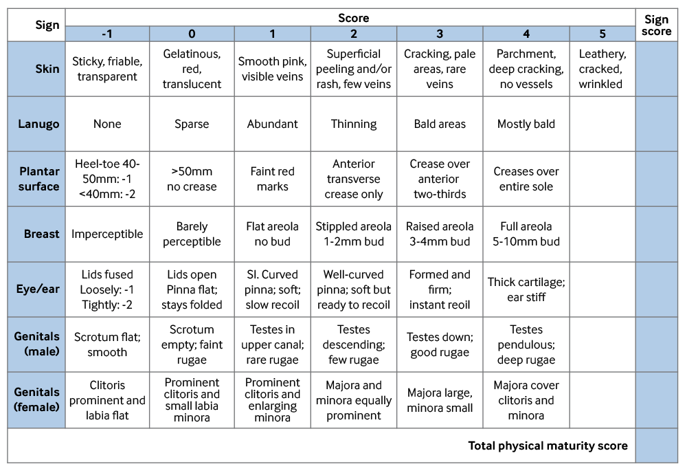
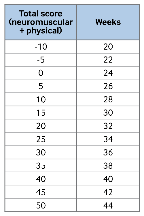
Postnatal methods of determining gestational age in premature infants, such as the New Ballard Score, have been developed and validated.[31] These assessments use measurements of neuromuscular and physical maturity to complement the historical or ultrasonographic estimates. The physical maturity scoring must be combined with the neuromuscular maturity scoring to assess correct gestational age. [Figure caption and citation for the preceding image starts]: Physical maturity, New Ballard Scoring SheetFrom Journal of Pediatrics 1991;119:417-423; used with permission [Citation ends]. [Figure caption and citation for the preceding image starts]: Neuromuscular maturity, New Ballard Scoring SheetFrom Journal of Pediatrics 1991;119:417-423; used with permission [Citation ends].
 [Figure caption and citation for the preceding image starts]: Maturity rating, New Ballard Scoring SheetFrom Journal of Pediatrics 1991;119:417-423; used with permission [Citation ends].
Use of this content is subject to our disclaimer