Investigations
1st investigations to order
chest x-ray: posterior-anterior (PA) and lateral
Test
Less sensitive than a CT scan and more specific than pulmonary function testing.
Left and right oblique views can increase sensitivity in comparison to PA view to identify pleural changes.
The presence of interstitial fibrosis in the lower zones and bilateral pleural thickening is highly specific.[1][2][Figure caption and citation for the preceding image starts]: Posterior-anterior view of the chest with bibasilar linear interstitial changes consistent with asbestosisFrom the personal collection of Kenneth D. Rosenman MD [Citation ends].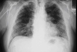 [Figure caption and citation for the preceding image starts]: Posterior-anterior view of the chest with 'en face' pleural changes in the mid zones on the right and left (arrows)From the personal collection of Kenneth D. Rosenman MD [Citation ends].
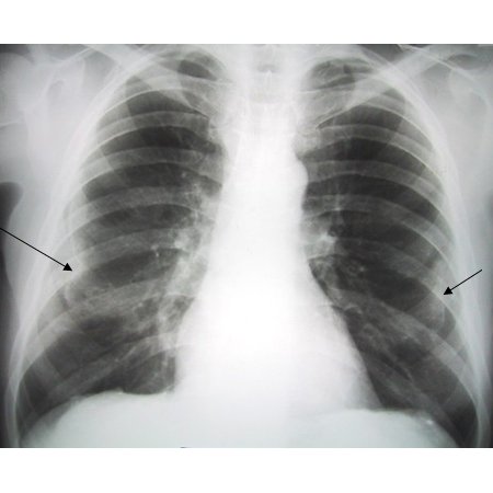 [Figure caption and citation for the preceding image starts]: Diffuse pleural thickening (arrowheads) and elevated left hemidiaphragm (dotted arrow)BMJ Case Reports 2009; doi:10.1136/bcr.06.2008.0253 [Citation ends].
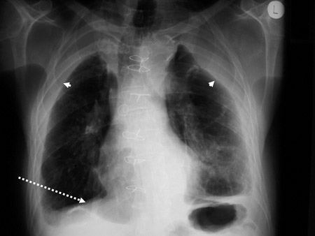 [Figure caption and citation for the preceding image starts]: Posterior-anterior view of the chest with 'mesa'-like pleural thickening of the left diaphragm and 'in-profile' pleural thickening of the mid zones of both the left and right lungsFrom the personal collection of Kenneth D. Rosenman MD [Citation ends].
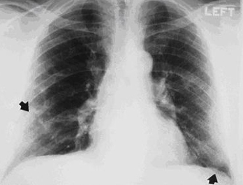
Result
lower zone linear interstitial fibrosis; progressively involves the entire lung; pleural thickening
pulmonary function tests
Test
Non-specific. The forced expiratory volume at 1 second (FEV₁) and forced vital capacity (FVC) can be used to demonstrate airflow limitation.
Restrictive: FVC, normal FEV1/FVC ratio, reduced slow vital capacity (SVC), reduced total lung capacity (TLC), reduced lung diffusion capacity testing (DLCO).
Obstructive changes: reduced FEV1, reduced FEV1/FVC ratio, increased residual volume (RV)/TLC ratio, reduced DLCO.
May have mixed restrictive and obstructive changes.
Reduced DLCO is the most sensitive pulmonary function change.[1]
Perform pulmonary function tests following standard guidelines.[16][17]
Result
restrictive changes; may have obstructive picture (especially if history of asbestos exposure and smoking)
Investigations to consider
high-resolution CT chest
Test
High-resolution CT (HRCT) chest scan is more sensitive than chest x-ray.[1][Figure caption and citation for the preceding image starts]: CT scan of the chest showing multiple examples of pleural thickening most with calcification (arrows)From the personal collection of Kenneth D. Rosenman MD [Citation ends].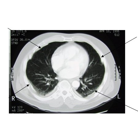 [Figure caption and citation for the preceding image starts]: CT scan confirming symmetrical thickening (arrowheads) with a calcified pleural plaque (broken arrow, top right) and an area of rounded atelectasis (Blesovsky's sign; dotted arrow, bottom right)Adapted from BMJ Case Reports 2009; doi:10.1136/bcr.06.2008.0253 [Citation ends].
 An HRCT scan should be performed if the individual has shortness of breath not explained by the chest x-ray or pulmonary function test results.
Result
lower zone linear interstitial fibrosis; progressively involves the entire lung; pleural thickening
lung biopsy
Test
Open lung biopsy is rarely needed for diagnosis. Its use should be limited to when cancer is suspected or in the absence of a known history of exposure to asbestos.
Bronchoscopic biopsy generally provides insufficient tissue to rule out asbestosis and is limited to evaluation for cancer, or if other clinical conditions are suspected.
Quantification of the mineral content is the most sensitive procedure.
Result
interstitial fibrosis; pleural changes with asbestos bodies; increased parenchymal asbestos mineral fibres
bronchial lavage
Test
Asbestos bodies may be found in bronchial lavage fluid and their presence is diagnostic of significant exposure and a high likelihood of asbestos-related disease and or radiographical changes.[21] However, asbestos bodies are unusual and their absence cannot be used to rule out asbestosis. Given this, bronchoscopic lavage has little clinical usage in evaluating a patient with suspected asbestosis, except in a research setting.
Result
presence of asbestos bodies in lavage fluid
Use of this content is subject to our disclaimer