Images and videos
Images

Asbestosis
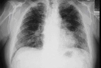
Posterior-anterior view of the chest with 'mesa'-like pleural thickening of the left diaphragm and 'in-profile' pleural thickening of the mid zones of both the left and right lungs
From the personal collection of Kenneth D. Rosenman MD
See this image in context in the following section/s:
Asbestosis
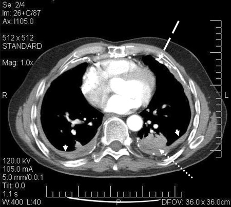
CT scan confirming symmetrical thickening (arrowheads) with a calcified pleural plaque (broken arrow, top right) and an area of rounded atelectasis (Blesovsky's sign; dotted arrow, bottom right)
Adapted from BMJ Case Reports 2009; doi:10.1136/bcr.06.2008.0253
See this image in context in the following section/s:
Asbestosis
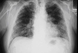
Posterior-anterior view of the chest with bibasilar linear interstitial changes consistent with asbestosis
From the personal collection of Kenneth D. Rosenman MD
See this image in context in the following section/s:
Asbestosis
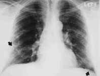
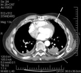
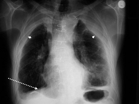
Diffuse pleural thickening (arrowheads) and elevated left hemidiaphragm (dotted arrow)
BMJ Case Reports 2009; doi:10.1136/bcr.06.2008.0253
See this image in context in the following section/s:
Asbestosis
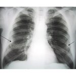
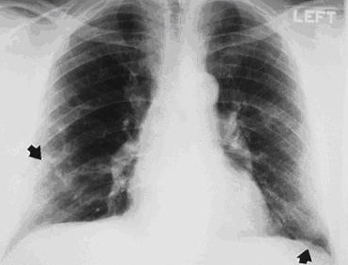
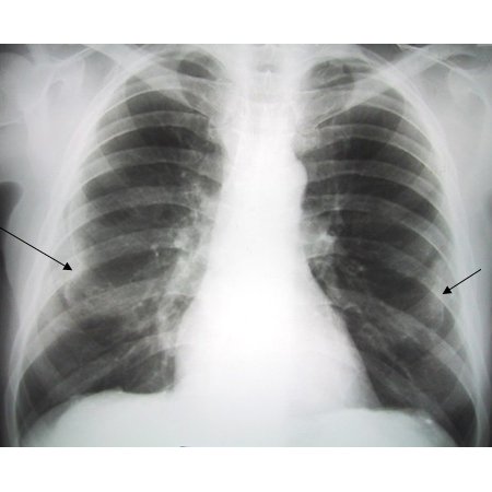
Posterior-anterior view of the chest with 'en face' pleural changes in the mid zones on the right and left (arrows)
From the personal collection of Kenneth D. Rosenman MD
See this image in context in the following section/s:
Asbestosis
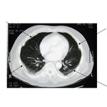
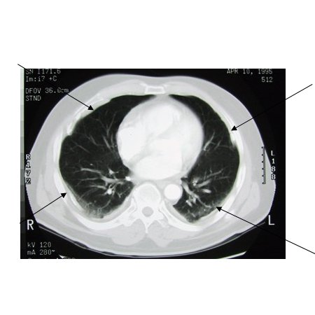
CT scan of the chest showing multiple examples of pleural thickening most with calcification (arrows)
From the personal collection of Kenneth D. Rosenman MD
See this image in context in the following section/s:
Use of this content is subject to our disclaimer