Differentials
Photo-allergic reaction
SIGNS / SYMPTOMS
History of potential photo-allergic substances, both topical (e.g., ultraviolet radiation (UVR) filters, such as PABA [para-aminobenzoic acid], cinnamates, artificial scents) and systemic medicines, such as antidepressants, antihypertensive medicine, antiphlogistics, or antibiotics.
Typical clinical signs include irregular erythema, papulovesicles, infiltration with scaling, and crusts.
The clinical manifestation resembles that of an allergic contact dermatitis, but confined to UVR-exposed areas. Some dissemination may occur. The typical symptom is itching. [Figure caption and citation for the preceding image starts]: Photo-allergic dermatitis after administration of a new diureticImage provided by Dr Hadshiew; used with permission [Citation ends].
INVESTIGATIONS
Photo-patch testing demonstrates a delayed reaction, with a slowly increasing (crescendo) inflammatory/eczematous reaction.
Phototoxic reaction
SIGNS / SYMPTOMS
Typical clinical signs are an amplified sunburn reaction and a positive history for phototoxic agents, both topical (e.g., tar, psoralens, giant hogweed, certain perfumes) and systemic medicines, such as antidepressants, diuretics, antiphlogistics, psoralens, or antibiotics (especially, tetracycline).
Typical symptoms are stinging and burning, even pain. [Figure caption and citation for the preceding image starts]: Phototoxic dermatitis after application of a new perfumeImage provided by Dr Hadshiew; used with permission [Citation ends].
In PLE there is usually only itching.
INVESTIGATIONS
Photo-patch testing demonstrates a sharply delineated, immediate, or delayed reaction, with a slowly decreasing (decrescendo) reaction.
Chronic actinic dermatitis
SIGNS / SYMPTOMS
Persistent light reaction, actinic reticuloid, or chronic photosensitive dermatitis are synonyms.[46][52][53]
Typically UV-B-induced (while PLE is more commonly UV-A-induced). Symptoms evolve over several days (like PLE), but persist for many weeks and form thick plaques, eczematous lesions with scaling and erosions, and lichenification. [Figure caption and citation for the preceding image starts]: Severe chronic actinic dermatitisImage provided by Dr Hadshiew; used with permission [Citation ends].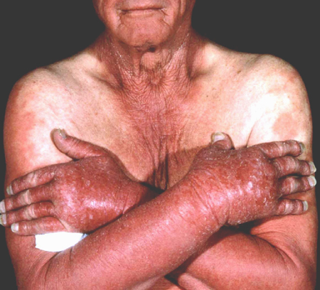
INVESTIGATIONS
Single photoprovocation on previously photoprotected skin causes induction of a persistent dermatitis (within 72 hours).
Skin biopsy and histopathological examination show spongiotic changes in the epidermis with uneven epidermal hyperplasia, crusts, and individual necrotic keratinocytes. The dermo-epidermal junction shows vacuolar alteration accompanied by a pronounced perivascular lymphocytic and sometimes also eosinophilic infiltration around the superficial and deep plexus.
Solar urticaria
SIGNS / SYMPTOMS
Clinical signs consist of urticaria on ultraviolet radiation-exposed sites that disappear quickly. The urticarial variant of PLE might resemble this presentation; however, skin lesions are visible for a few days in PLE.
INVESTIGATIONS
Single photoprovocation on previously photoprotected skin demonstrates immediate (up to 1 hour) induction of urticarial patches. Visible or infrared light might be part of the induction spectrum. If conventional tests are negative, exposure to natural sunlight may be considered.[54]
Erythema exudativum multiforme
SIGNS / SYMPTOMS
Photosensitive variants of erythema exudativum multiforme are rare but difficult to distinguish from the erythema exudativum multiforme type of PLE.[4] Typically, a concurrent infection with herpes simplex virus is present together with intensive ultraviolet radiation-exposure, which elicits symptoms.[4]
INVESTIGATIONS
Diagnosis is clinical.
Systemic lupus erythematosus
SIGNS / SYMPTOMS
Typical signs involve the delayed appearance of erythematous plaques on ultraviolet radiation (UVR) exposed sites that persist over a prolonged period of time (weeks).
Typical symptoms are stinging and burning. Skin lesions disappear within a few days (after avoiding UVR exposure). [Figure caption and citation for the preceding image starts]: Typical butterfly (or malar) rash in lupus erythematosusImage provided by Dr Hadshiew; used with permission [Citation ends].
INVESTIGATIONS
Repetitive photoprovocation on previously photoprotected predilection sites (upper arms, shoulders). Induction of typical skin lesions might take time (several days up to 3 weeks) and require several weeks for remission.[55]
Skin biopsy and histopathological examination with immunofluorescence shows atrophic changes in the epidermis with vacuolar degeneration, band-like infiltration of immunoglobulins, and complement along the basal membrane (positive direct immunofluorescence; absent in PLE).
ANA, anti-Ro (SS-A), and anti-La (SS-B) tests should be obtained. In PLE they are negative.
Lymphocytic infiltration
SIGNS / SYMPTOMS
Clinical signs include persistent plaques and papules, similar to PLE.[56] Signs persists longer than PLE.
INVESTIGATIONS
Repetitive photoprovocation on previously photoprotected predilection sites leads to the induction of typical skin lesions within a few days.
Histopathological examination shows similar signs to PLE, but with no sub-epidermal oedema.
Fixed sunlight eruption
SIGNS / SYMPTOMS
Three case studies have described a condition similar to fixed drug eruption, caused by exposure to UV-A and UV-B, as opposed to medications.[57][58][59] Signs include round and well-demarcated macules, plaques, or patches of hyperpigmentation or erythema. These are relatively persistent and localised. Patients experience a burning sensation; however, there are no reports of vesicles or papules in cases of fixed sunlight eruption.
INVESTIGATIONS
Histology reveals characteristics similar to those of fixed drug eruptions, such as spongiosis and apoptotic keratinocytes.
Use of this content is subject to our disclaimer