Differentials
Hip dysplasia
SIGNS / SYMPTOMS
Caused by intrauterine loss of contact between the fetus' developing femoral head and acetabulum.
May range from mild, asymptomatic cases to severe dysplasia with congenital hip dislocation.
Moderate to severe dysplasia may predispose to early osteoarthritis, labral tear, or impingement, and present with findings secondary to 1 or more of these conditions.
Patients with congenital dislocation will have shortening of the involved leg and decreased hip range of motion.
INVESTIGATIONS
Shallow, more vertically oriented acetabulum seen on plain films.
Nerve entrapment
SIGNS / SYMPTOMS
Rare cause of groin pain.
Obturator neuropathy may result in exercise-induced groin pain, relieved by rest. There may be discernible adductor muscle weakness on motor examination if the patient's symptoms are present, and decreased sensation along the medial thigh.
Lateral femoral cutaneous nerve entrapment or "meralgia paraesthetica" typically causes a patch of numbness and/or burning pain along the anterolateral thigh. There is no associated motor weakness with this condition.
Ilio-inguinal nerve compression may cause pain in the groin or genital region. It is most commonly seen in patients with a history of prior abdominal surgery or hypertrophic abdominal muscles.
INVESTIGATIONS
Diagnosis of obturator nerve entrapment can be confirmed by EMG that shows chronic denervation changes along the affected muscle group.
Diagnosis of lateral femoral cutaneous or ilio-inguinal nerve entrapment may be made by local infiltration of lidocaine anaesthetic, which should relieve symptoms within minutes.
Ankylosing spondylitis
SIGNS / SYMPTOMS
Spondyloarthropathy typically seen in young to middle-aged men.
Hip joint involvement is common, often bilateral.
Symptoms (stiffness, pain) are worse in the morning and improve during the course of the day.
Low back and sacro-iliac joints are also frequently affected.
INVESTIGATIONS
Plain film x-rays of the hip and pelvis may demonstrate irregularities of the sacro-iliac joints with possible erosive and sclerotic changes.
The lumbar spine may reveal bridging syndesmophytes of multiple vertebrae leading to the classic "bamboo spine" appearance.
Rheumatoid arthritis
SIGNS / SYMPTOMS
Systemic inflammatory disease with multiple joint involvement.
While classically associated with small joints of the hands and feet, hip joint involvement is commonly seen.
Monoarticular involvement of the hip joint as a presenting symptom in an undiagnosed rheumatoid patient, while rare, can be mistaken for septic arthritis due to the marked inflammatory process and joint synovitis.
INVESTIGATIONS
Plain film x-rays may demonstrate varying degrees of concentric joint space narrowing, cysts, and periarticular erosions. Unlike in osteoarthritis, osteophytes and sclerotic changes are not typically present.
Definitive serological tests, such as rheumatoid factor or anti-citrullinated protein antibodies (ACPA), are positive in around 70% of patients.[20]
Osteoarthritis
SIGNS / SYMPTOMS
Typically seen in patients aged >50 years. Presents with activity-related pain; in advanced cases there may be a component of rest or night pain. Limited range of motion with pain at the extremes (especially flexion and internal rotation). Hip flexion contracture may be present. Positive Stinchfield's and/or Trendelenburg's sign.
INVESTIGATIONS
Plain film radiographs with anteroposterior (AP) pelvis, AP and lateral hip, or frog's leg view may demonstrate joint space narrowing, sclerotic and/or cystic changes, and osteophyte formation.
Osteonecrosis
SIGNS / SYMPTOMS
History of recent trauma (femoral neck fracture, hip dislocation) or presence of risk factors (excessive alcohol consumption, chronic corticosteroid use). Bilateral in up to 50% of cases.
INVESTIGATIONS
Plain film radiographs may be unremarkable in early stage of disease. Later stages characterised by subchondral collapse with formation of a "crescent sign".
MRI is the definitive test in these circumstances and may demonstrate bony oedema, revascularisation changes seen early in the disease process.
Lumbar disc pathology
SIGNS / SYMPTOMS
Rare cause of groin pain. Upper lumbar disc herniation (L1 or L2) may cause referred pain to groin area.
Pain typically worse with Valsalva manoeuvres (e.g., sneezing or bearing down). Back and buttock pain may be present.
Hip examination is unremarkable. Motor examination may demonstrate hip flexor weakness. Sensation may be subjectively decreased along dermatomal distribution.
INVESTIGATIONS
Plain film x-rays of lumbar spine may demonstrate degenerative disc disease of upper lumbar vertebrae.
MRI of the lumbar spine should be obtained to rule out disc herniation or foraminal narrowing impinging on corresponding lumbar nerve root.
Septic arthritis
SIGNS / SYMPTOMS
Acute presentation. Joint is usually hot, swollen, and tender, with restricted movement, severe pain on weight-bearing. Fever may be present. More common in joints with a prosthesis. Resting position of hip flexion, abduction, and external rotation may be adopted to relieve pain.
INVESTIGATIONS
FBC with differential shows raised WBC count. Blood cultures may be positive. ESR and C-reactive protein elevated. Synovial fluid Gram stain and WBC count positive.
Osteomyelitis
SIGNS / SYMPTOMS
Typically a chronic or acute-on-chronic presentation, with vague pain complaints. May have rest or night pain. Constitutional symptoms (fevers, chills, malaise) often present. An unremarkable hip examination possible if the infectious process involves the pelvis.
INVESTIGATIONS
FBC with differential may show elevated WBC count. Blood cultures may be positive. ESR and C-reactive protein may be elevated.
Plain film radiographs may show changes consistent with chronic osteomyelitis in some cases. MRI has higher sensitivity and specificity: up to 98% and 89%, respectively.[21]
Bone malignancy or metastases
SIGNS / SYMPTOMS
Typically results in severe pain, which is often worse at night.
INVESTIGATIONS
CT/MRI may reveal the metastatic lesion. [Figure caption and citation for the preceding image starts]: Metastatic lesion of the femoral neck seen on CTFrom the collection of Cedric J. Ortiguera, MD [Citation ends].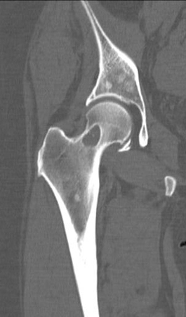
Congenital or anatomical deformity
SIGNS / SYMPTOMS
Common degenerative conditions, such as osteoarthritis, or anatomical and mechanical distortions seen in conditions such as femoroacetabular impingement or hip dysplasia, may cause groin pain.
INVESTIGATIONS
Radiographic findings suggestive of congenital or anatomical deformity. Patients with radiographic findings that suggest a congenital or anatomical deformity should be referred to an orthopaedic surgeon for evaluation and treatment recommendations.
Referred pain syndromes
SIGNS / SYMPTOMS
May be referred from non-musculoskeletal areas, including the gastrointestinal tract (e.g., appendicitis, diverticulosis, inflammatory bowel disease), genitourinary tract (e.g., urinary tract infection, prostatitis, nephrolithiasis) and neuroradicular (e.g., sacroiliac joints, lumbar disk pathology).
INVESTIGATIONS
Tests depend on the source of the pain.
Use of this content is subject to our disclaimer