Case history
Case history #1
A 32-year-old man who plays football 2 or 3 times a week presents with groin pain that has been present for the past 3 to 4 months. At first it occurred only after sport, but it has gradually got worse and now he cannot play any more. He has pain when sprinting and turning and when kicking the ball, and after playing he has pain for 2 to 3 days. He also has pain when sneezing and coughing. No relevant trauma can be recollected.
Case history #2
A 40-year-old woman who runs 5 to 6 kilometres (3 to 4 miles), 3 times a week, presents after noticing increased pain in the groin during a 2-week hill-walking holiday. Since the holiday, she has had pain after her regular running, after sitting in a deep chair for a while, and when climbing stairs. Sometimes she also has slight low back pain.
Other presentations
Acute-onset hip or groin pain is often accompanied by a history of recent trauma, sport-related injury, or overuse. Groin pain in the endurance or recreational athlete who has increased his/her level of activity should be assessed radiographically for possible stress fracture. Patients with septic arthritis (more common in the paediatric population) present with difficulty weight-bearing on the involved extremity and pain with passive range of motion of the joint. Accompanying clinical signs may include fever. Groin pain that is aggravated by Valsalva manoeuvre may be due to lumbar disc pathology or femoral/inguinal herniation. In patients with ill-defined pain or systemic symptoms such as fevers, nausea, and anorexia, intra-abdominal pathology should be suspected. Chronic groin pain or groin pain with atypical features, such as rest or night pain and/or a history of cancer, warrants x-ray evaluation to rule out malignancy. [Figure caption and citation for the preceding image starts]: MRI demonstrating inferior right femoral neck stress fracture (compression-sided)From the collection of Cedric J. Ortiguera, MD [Citation ends].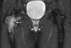 [Figure caption and citation for the preceding image starts]: Metastatic lesion of the femoral neck seen on CTFrom the collection of Cedric J. Ortiguera, MD [Citation ends].
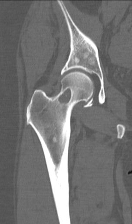
Use of this content is subject to our disclaimer