Images and videos
Images


Syphilis infection
This was a case of congenital syphilis resulting in the death of this newborn infant
CDC: PHIL image ID 3510; used with permission
See this image in context in the following section/s:
Syphilis infection
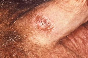
A penile chancre located on the proximal penile shaft: primary syphilitic infection
CDC/ Dr Gavin Hart; Dr NJ Fiumara; used with permission
See this image in context in the following section/s:
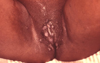
Syphilis infection
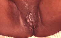
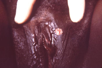
A primary vulvar syphilitic chancre due to Treponema pallidum bacteria
CDC: PHIL image ID 5340; used with permission
See this image in context in the following section/s:
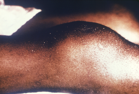
Syphilis infection
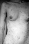
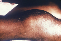
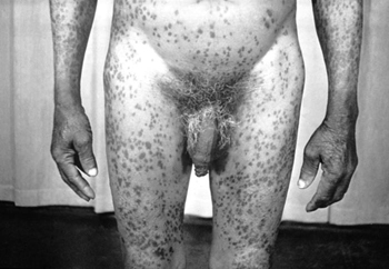
Secondary syphilis presenting pigmented macules and papules on the skin
CDC/Susan Lindsley; used with permission
See this image in context in the following section/s:


Syphilis infection
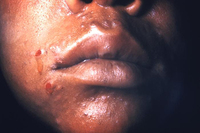
Secondary syphilitic lesions on the face
CDC: PHIL image ID 3500; used with permission
See this image in context in the following section/s:
Syphilis infection
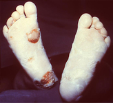
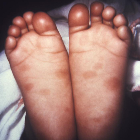
This newborn presented with symptoms of congenital syphilis that included lesions on the soles of both feet
CDC: PHIL image ID 4148; used with permission
See this image in context in the following section/s:
Syphilis infection
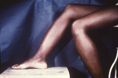
Osteoperiostitis of the tibia ('saber shins')
CDC/Robert E. Sumpter; used with permission
See this image in context in the following section/s:
Syphilis infection
Secondary syphilitic lesions of vagina
CDC/J. Pledger; used with permission
See this image in context in the following section/s:

Syphilis infection
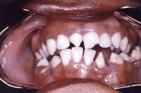
Peg-shaped, notched central incisors (Hutchinson's teeth)
CDC/Robert E. Sumpter; used with permission
See this image in context in the following section/s:
Syphilis infection
Clutton's joints
CDC/Richard Deitrick; used with permission
See this image in context in the following section/s:
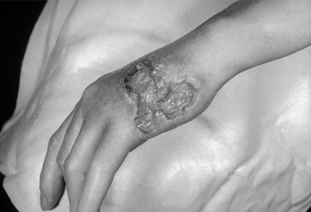
Syphilis infection
Gummatous lesions on the dorsal surface of the left hand
CDC/Susan Lindsley; used with permission
See this image in context in the following section/s:

Syphilis infection
Secondary syphilitic papulosquamous rash on the torso and upper body
CDC/Susan Lindsley; used with permission
See this image in context in the following section/s:
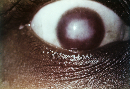
Syphilis infection
Interstitial keratitis
CDC/Susan Lindsley
See this image in context in the following section/s:
Videos
 Diagnostic lumbar puncture in adults: animated demonstration
How to perform a diagnostic lumbar puncture in adults. Includes a discussion of patient positioning, choice of needle, and measurement of opening and closing pressure.
Use of this content is subject to our disclaimer