Case history
Case history #1
A 27-year-old man notes a painless penile ulcer. He has recently started a new relationship. He is otherwise asymptomatic, as is his partner. On examination, the ulcer is indurated and the inguinal lymph nodes are rubbery and moderately enlarged.
Case history #2
A 30-year-old man presents with difficulty hearing conversations while in a crowded room. Following referral for audiometry, bilateral high-frequency hearing loss is diagnosed. On further questioning he reports a past history of an anal fissure about 10 weeks previously that healed spontaneously. He also describes a mild transient skin rash 2 weeks before his auditory symptoms appeared. He says that he has been feeling unusually tired.
Other presentations
The chancre (syphilitic ulcer) of primary syphilis is usually a solitary, painless, clean ulcer with an indurated base. Less commonly, there may be multiple or painful ulcers. This presentation may represent co-infection with chancroid or genital herpes. Uncommon presentations of secondary syphilis include specific organ involvement, such as hepatitis, iritis, chorioretinitis, glomerulonephritis, nephrotic syndrome, neurosyphilis involving cranial nerves (particularly the eighth cranial nerve), and meningitis.[9] Gummata are an extremely rare manifestation of tertiary (late) syphilis. Gummatous syphilis (also known as benign tertiary syphilis) usually affects skin and bone but can also affect visceral organs, causing organomegaly or infiltrative or destructive lesions, as well as perforation or collapse of affected structures.[4][9]
[Figure caption and citation for the preceding image starts]: A primary vulvar syphilitic chancre due to Treponema pallidum bacteriaCDC: PHIL image ID 5340; used with permission [Citation ends].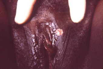 [Figure caption and citation for the preceding image starts]: A penile chancre located on the proximal penile shaft: primary syphilitic infectionCDC/ Dr Gavin Hart; Dr NJ Fiumara; used with permission [Citation ends].
 [Figure caption and citation for the preceding image starts]: Gummatous lesions on the dorsal surface of the left handCDC/Susan Lindsley; used with permission [Citation ends].
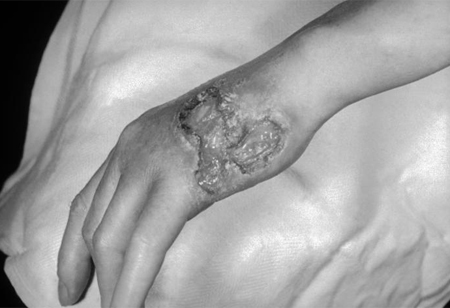
Use of this content is subject to our disclaimer