Investigations
1st investigations to order
CT head scan
Test
Preferred initial diagnostic study when subarachnoid haemorrhage is suspected.
About 95% of patients have evidence of subarachnoid blood on a non-contrast head CT obtained within the first 48 hours after rupture.
Result
subarachnoid blood in ruptured or leaking aneurysm; calcified or thrombosed aneurysm may also be seen on CT
conventional catheter-based angiogram
Test
Highest spatial resolution in determining aneurysm size, location, and morphology in relation to nearby arteries.
Three-dimensional reconstruction allows for further resolution of aneurysm configuration.
Initial test after ruptured aneurysm has been diagnosed on CT or lumbar puncture.[Figure caption and citation for the preceding image starts]: Cerebral angiogram showing aneurysmFrom the personal collection of Dr M. Chen, Columbia College of Physicians and Surgeons [Citation ends].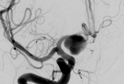 [Figure caption and citation for the preceding image starts]: Comparison of 2-dimensional catheter angiography (left) with 3-dimensional catheter angiography (right) showing a basilar tip aneurysmFrom: Sellar M. Practical Neurology. 2005;5:28-37. Used with permission [Citation ends].
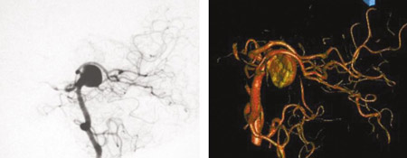 [Figure caption and citation for the preceding image starts]: Three-dimensional catheter angiogram showing a basilar tip aneurysmFrom: Sellar M. Practical Neurology. 2005;5:28-37. Used with permission [Citation ends].
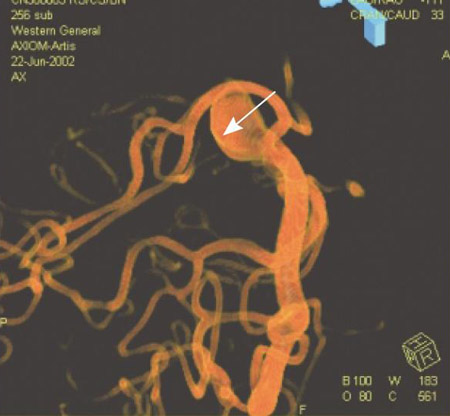
Result
aneurysm in relation to arteries
CT angiography
magnetic resonance angiography (MRA)
Test
Takes longer to perform than CT angiography, therefore less appropriate for critically ill patients. High specificity in detecting aneurysms >3 mm.[27][29]
MRA does not carry the risk of ionising radiation and can be obtained without contrast using time of flight techniques (MRA-TOF).
Can be used as an initial test for unruptured cerebral aneurysms and is the investigation of choice for aneurysm screening.
Result
aneurysm location/size
Investigations to consider
lumbar puncture
Test
Contraindicated if there is a suspicion for a mass lesion (lateralising neurological deficits or papilloedema).
Reserved for about 5% of patients in whom head CT is normal with suspected subarachnoid haemorrhage. It is reasonable to exclude SAH if a scan performed within 6 hours does not show subarachnoid blood.[20] However, if there is high clinical suspicion for SAH but the CT scan was performed after 6 hours, a lumbar puncture should be performed.[20][21]
Bloody cerebrospinal fluid (CSF) that does not clear with continued egress of fluid raises index of suspicion.
The presence of xanthochromia, a yellowish discoloration of CSF representing bilirubin, is more specific than elevated red cell count in CSF.
Result
elevated red blood cell count with xanthochromia
Use of this content is subject to our disclaimer