Images and videos
Images


Cerebral aneurysm
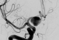
Cerebral angiogram showing aneurysm
From the personal collection of Dr M. Chen, Columbia College of Physicians and Surgeons
See this image in context in the following section/s:

Cerebral aneurysm
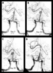
Progressive angiography images of a small dissecting aneurysm of the distal basilar artery after a subarachnoid and intraventricular haemorrhage on day 3 (A), day 23 (B), and day 30 (C), and 6 months after stent-assisted coiling (D). Arrows indicate proximal and distal stent markers
From: Peluso JP, van Rooij WJ, Sluzewski M. BMJ Case Reports 2009; doi:10.1136/bcr.2007.121533. Used with permission
See this image in context in the following section/s:
Cerebral aneurysm
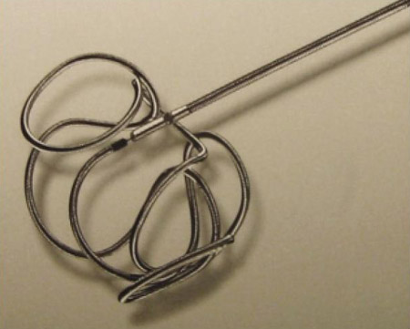
Example of a coil used to treat cerebral aneurysms
From: Sellar M. Practical Neurology. 2005;5:28-37. Used with permission
See this image in context in the following section/s:

Cerebral aneurysm
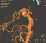
Three-dimensional catheter angiogram showing a basilar tip aneurysm
From: Sellar M. Practical Neurology. 2005;5:28-37. Used with permission
See this image in context in the following section/s:
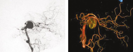
Cerebral aneurysm
Comparison of 2-dimensional catheter angiography (left) with 3-dimensional catheter angiography (right) showing a basilar tip aneurysm
From: Sellar M. Practical Neurology. 2005;5:28-37. Used with permission
See this image in context in the following section/s:
Use of this content is subject to our disclaimer