Images and videos
Images

Tricuspid stenosis
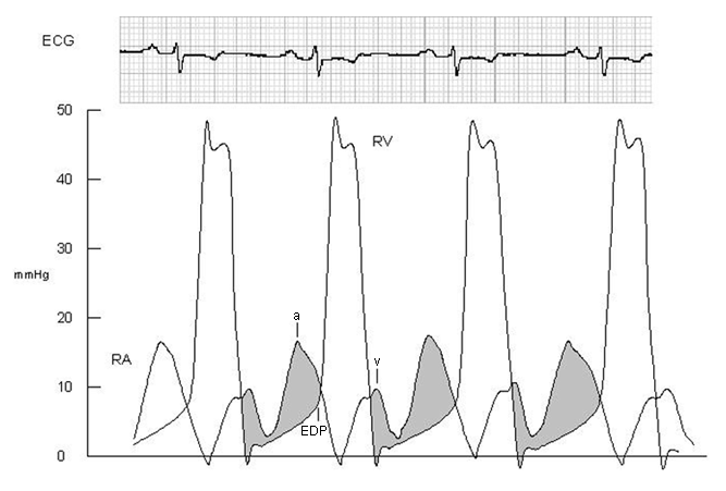
Haemodynamic tracings obtained during cardiac catheterisation from a woman with moderate to severe rheumatic tricuspid valve stenosis
From the personal collection of Martin Bocks; used with permission
See this image in context in the following section/s:

Tricuspid stenosis
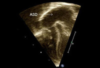
Congenital tricuspid valve stenosis: 2D transthoracic echocardiogram apical 4 chamber view reveals a small tricuspid valve annulus (dash), RV hypoplasia, and a large atrial septal defect (ASD, arrow)
From the personal collection of Martin Bocks; used with permission
See this image in context in the following section/s:
Tricuspid stenosis
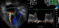
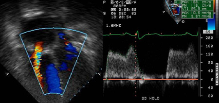
Echocardiogram with colour Doppler reveals flow acceleration across the tricuspid valve and spectral Doppler reveals a mean TV gradient of 6 mmHg
From the personal collection of Martin Bocks; used with permission
See this image in context in the following section/s:
Use of this content is subject to our disclaimer