Images and videos
Images


Assessment of hearing loss
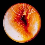
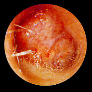
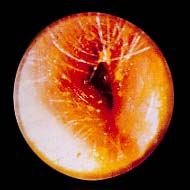
Erythematous bulging tympanic membrane due to acute otitis media
From the collection of Dr Richard Buckingham
See this image in context in the following section/s:
Assessment of hearing loss
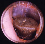
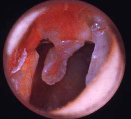
Squamous cell carcinoma of the ear canal
From the collection of Dr Richard Buckingham
See this image in context in the following section/s:
Assessment of hearing loss
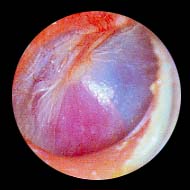
Right tympanic membrane perforation
From the collection of Dr Richard Buckingham
See this image in context in the following section/s:
Assessment of hearing loss
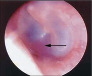
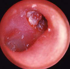
Right attic cholesteatoma
From the collection of Dr Richard Buckingham
See this image in context in the following section/s:
Assessment of hearing loss
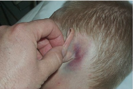
Battle’s sign
Reproduced with permission from van Dijk GW. The bare essentials: head injury. Pract Neurol. 2011 Feb;11(1):50-5.
See this image in context in the following section/s:
Assessment of hearing loss
Right ear with glomus tumour visible inferiorly behind intact tympanic membrane
From the collection of Dr Richard Buckingham
See this image in context in the following section/s:

Assessment of hearing loss
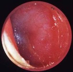
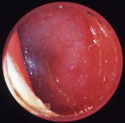
Haemotympanum
Reproduced with permission from van Dijk GW. The bare essentials: head injury. Pract Neurol. 2011 Feb;11(1):50-5.
See this image in context in the following section/s:
Assessment of hearing loss
Anatomy of the ear
Created by BMJ Knowledge Centre
See this image in context in the following section/s:
Assessment of hearing loss
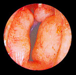
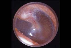
Picture of a normal left ear
From the collection of Dr Richard Buckingham
See this image in context in the following section/s:

Assessment of hearing loss
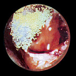
Otomycosis: fungal debris in ear canal
From the collection of Dr Richard Buckingham
See this image in context in the following section/s:
Assessment of hearing loss
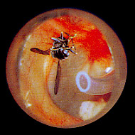
Insect in ear canal with pressure equalisation tube visible anteriorly
From the collection of Dr Richard Buckingham
See this image in context in the following section/s:
Assessment of hearing loss
Swollen ear canal, almost completely closed due to acute otitis externa
From the collection of Dr Richard Buckingham
See this image in context in the following section/s:

Assessment of hearing loss
Foreign body in ear canal
From the collection of Dr Richard Buckingham
See this image in context in the following section/s:

Assessment of hearing loss
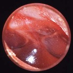
Left ear with effusion behind intact tympanic membrane
From the collection of Dr Richard Buckingham
See this image in context in the following section/s:

Assessment of hearing loss
Ear canal with bony narrowing secondary to exostoses
From the collection of Dr Richard Buckingham
See this image in context in the following section/s:
Videos
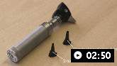 How to examine the ear
How to perform an examination of the ear.
Use of this content is subject to our disclaimer