Approach
A thorough history and physical examination are essential to reach the correct diagnosis. Hearing loss can be an isolated symptom or associated with other aural symptoms. Patients may or may not be aware that their hearing is decreased. They may state that their ear feels muffled, blocked, or plugged, or complain of pressure or the sensation of water in the ear.
The general approach is to decide if the hearing loss is due to a problem in the outer, middle, or inner ear. Problems in the outer and middle ear cause a conductive hearing loss and those in the inner ear cause a sensorineural hearing loss. It is possible that more than one problem may contribute to hearing loss.
History
Important questions include when onset of hearing loss occurred, whether the loss was acute or gradual, and whether there are any associated symptoms, such as ear pain, drainage of fluid from the ear, tinnitus, dizziness, vertigo, facial nerve weakness, or headache.
External ear
Gradual hearing loss in a patient with a hearing aid or in someone who admits to cleaning their ears with cotton swabs may be due to cerumen impaction. There may be associated tinnitus and/or pain depending on the degree of impaction.
In a small child with a foreign body in the external canal, there is often a clear history of putting something in the ear. If not witnessed, a delayed presentation with pain and infection may result.
Pain and swelling of the external canal may follow local trauma or recent instrumentation, such as syringing.
Recurrent ear infections with gradual hearing loss may be the result of a mechanical obstruction in the outer-ear canal, such as a polyp, osteoma, or exostosis.
A history of regular immersion in cold water, with activities such as swimming and surfing, suggests exostosis.
Symptoms of pain and drainage from the ear canal, especially following recent intervention, suggest otitis externa. Patients who are immunocompromised are more at risk.
Pain that persists after apparent resolution of external signs wakes the patient from sleep suggests necrotising otitis externa.
Chronic pain and ear drainage, as well as a lack of response to local treatment, can indicate neoplastic change necessitating further investigation.
Middle ear
Acute otitis media commonly presents in children. Typical symptoms include pain, fever, muffled hearing, and occasionally swelling behind the ear. Otitis media with effusion (OME) may be suspected if, upon resolution of acute otitis media, the patient complains that their hearing is still decreased.[77][78] Patients with suspected OME may report a recent upper respiratory tract infection (URTI) or aeroplane travel.
Recurrent ear drainage with a strong odour and a history of chronic ear infections suggest cholesteatoma formation, particularly if there has been a tympanic membrane perforation in the past. There may also be associated symptoms of dizziness and tinnitus, although these are non-specific.
Eustachian tube dysfunction presents as a feeling of fullness in the affected ear, a popping sensation (as commonly experienced when descending in an aircraft), and sometimes with pain or dizziness. The symptoms may follow a recent URTI or with allergic rhinitis and may last up to 1 week.
A history of acute trauma, such as a blow to the head or barotrauma from scuba diving with sudden onset of hearing impairment, may suggest tympanic-membrane perforation. Scuba divers may report increasing pain with sudden relief as the tympanic membrane perforates to equalise the pressures on each side. A large perforation will enable cold water to enter the middle ear and this may cause feelings of nausea or disequilibrium.
The presence of bloody discharge from the ear or clear fluid from the nose following trauma, such as a blow to the head, may result from temporal bone fracture.
Inner ear
Sudden hearing loss with vertigo, nausea, and vomiting is likely to be due to labyrinthitis.
Recurrent episodes of vertigo with fluctuating hearing loss and tinnitus are characteristic of Meniere's disease.[35]
Previous exposure to sustained high levels of noise or sudden loud noises through occupational use of power tools or hobbies, such as motorbike racing or shooting, may cause gradual difficulty in understanding speech in loud environments.
Bilateral, gradual-onset hearing loss is commonly age related (presbycusis); patients aged >50 years may be screened for hearing loss at health care encounters.[32] In the younger adult with progressive bilateral hearing loss, otosclerosis should be considered. Tinnitus may or may not be present.
A perilymphatic fistula can be confirmed only by direct visualization at exploratory surgery but may be suspected by features in the history, such as fluctuating hearing loss with vertigo with a past history of surgery on the stapes or previous barotrauma.
Unilateral hearing loss, with associated symptoms of tinnitus, nausea, and facial weakness, strongly suggests the possibility of an acoustic neuroma. In patients with known neurofibromatosis, this is particularly likely, although their symptoms tend to be bilateral with acoustic neuromas on both sides.
A complaint of pulsatile tinnitus with hearing impairment and a feeling of fullness may indicate a rarer glomus tumour.
Hearing loss in children
Hearing loss can occur as an isolated genetic abnormality or as one feature of a genetic syndrome.
Some hereditary causes can develop later in childhood and, because they are often autosomal recessive, both parents may have entirely normal hearing.
A thorough prenatal and family history will be particularly useful in eliciting clues.
A severely jaundiced baby with apparent hearing loss may be experiencing the directly toxic effects of high levels of unconjugated bilirubin.[48][49]
Consider congenital infection (e.g., cytomegalovirus), genetic syndromes and previous meningitis infection in children who have hearing loss in conjunction with other neurological symptoms, such as seizures, spasticity, and developmental delay.
Many babies with hearing loss are now being discovered by newborn hearing screening, which allows for early detection and intervention.[79] Formal recommendations in the US for evaluation and referral of children with early identified hearing loss are available.[80]
Systemic illness
Several systemic illnesses can feature hearing loss as a complication, and this may occur in a patient with a known illness or, more rarely, as the presenting feature. However, other causes of hearing loss, such as cerumen impaction, noise-related hearing loss, and presbycusis, can be responsible, even in the presence of a systemic disease known to cause potential hearing loss.
Pain and deformity in other bones often occurs with Paget's disease and hearing loss can be unilateral or bilateral.
Systemic lupus erythematosus (SLE) and multiple sclerosis can cause a sudden hearing loss.[53][81]
Symptoms of rhinitis, nosebleeds, and haematuria may suggest underlying granulomatosis with polyangiitis (formerly known as Wegener's granulomatosis).
Gradual hearing loss can occur with osteogenesis imperfecta, commonly in the second to fourth decades of life.
Sudden unilateral hearing loss following minor neck trauma and the presence of an occipital headache should raise the possibility of a vertebral artery dissection.
Hearing loss with new symptoms of ataxia, speech difficulties, and/or hemiparesis is consistent with a stroke, particularly with a history of cardiovascular risk factors, such as hypertension and smoking.
Facial pain, headaches, and muscle weakness can be features of Arnold-Chiari syndrome, which is the gradual downwards displacement of the cerebellar tonsils.
Drug-related
Aminoglycoside antibiotics, such as gentamicin, azithromycin, nonsteroidal anti-inflammatory drugs, chemotherapeutic drugs, antimalarial drugs, and loop diuretics in high dose, can all be ototoxic.[33] Hearing loss as a result of ototoxic drugs usually starts at high frequencies. The effects are usually reversible if identified and stopped early, but hearing loss can become permanent if medication is continued so a high index of suspicion is required.
Physical examination
To determine whether the cause of hearing loss is conductive (outer or middle ear) or sensorineural (inner ear) requires a visual inspection of the outer-ear canal and tympanic membrane using an otoscope and performance of tuning fork tests. Removal of cerumen may be required to adequately examine the ear canal.[7] Any obstructing hairs or foreign bodies should also be removed to get a clear view of the tympanic membrane.
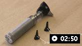
How to perform an examination of the ear.
Otoscopy
External ear
Ear-canal swelling (without trauma), granulation tissue, and yellowish otorrhoea suggest otitis externa. Fungal hyphae may be present. Persistent granulation tissue could be malignant. New bone growth along the upper part of the tympanic bone or squamotympanic membrane may indicate an osteoma (usually solitary). Multiple bony growths arising from the anterior and posterior parts of the external auditory canal may indicate exostosis.
Tympanic membrane
An erythematous, bulging tympanic membrane is consistent with acute otitis media.
If a red bulging mass can be seen behind the tympanic membrane, this could be a glomus tumour and a bruit can sometimes be heard.
Fluid visible behind the tympanic membrane, particularly when present following acute otitis media or upper respiratory tract infection, may suggest otitis media with effusion.[77][78] The tympanic membrane may appear normal, and there are no signs or symptoms of acute inflammation. There may be bubbles or an air-fluid level.[77]
Blood visible behind the tympanic membrane suggests a temporal bone fracture (with a consistent history).
A hole in the tympanic membrane confirms a perforation, either acutely or one that has not healed. A small perforation may be seen with gradual noise-related hearing loss. Tympanic-membrane perforations generally merit otolaryngology referral.
Necrotic debris may be seen in the middle ear and purulent drainage would raise suspicion of a cholesteatoma.
If the tympanic membrane appears normal but retracted, this means there is negative pressure in the middle ear and this may be due to Eustachian tube dysfunction preventing pressure equalisation.
A pneumatic otoscope can be used to check the mobility of the intact tympanic membrane:
A hypomobile tympanic membrane suggests middle-ear fluid or a fixed hypomobile ossicular chain (otosclerosis).
A hypermobile tympanic membrane can occur with ossicular discontinuity, which may follow a blow to the side of the head and is associated with temporal bone fracture.
Tympanometry can be used for patients who are difficult to examine and for patients in whom the diagnosis is uncertain following pneumatic otoscopy.[77]
Tuning fork tests
If the tympanic membrane is intact and has a normal appearance, then hearing loss may be secondary to pathology of the middle ear (conductive) or inner ear (sensorineural). Tuning fork testing can help to distinguish between them.[82]
Rinne's test: the tuning fork is placed on the mastoid bone (bone conduction) and then placed next to the external ear (air conduction). Usually air conduction is heard louder than bone conduction and this is a positive Rinne's test. If bone conduction is louder than air conduction, this is a negative Rinne's test and indicates conductive hearing loss in that ear.
Weber's test: the tuning fork is placed on the forehead. The patient is asked in which ear the sound is louder. If the patient hears the sound equally in each ear or cannot localise, this is normal and is termed a mid-line Weber's. The sound lateralises to the affected side in conductive hearing loss and to the unaffected side in sensorineural hearing loss. For example, if the patient hears the sound louder in the right ear, then this indicates either a right conductive or a left sensorineural hearing loss.
If a tuning fork is not available, ask the patient to hum. If the hum sounds louder in the affected ear, the patient likely has a conductive hearing loss in that ear.[2] The hum test has a sensitivity, specificity and diagnostic accuracy similar to the Weber test.[83]
Neurological examination
Cranial nerve palsies may occur in necrotising otitis externa, mastoiditis, cholesteatoma, temporal bone fracture, stroke, acoustic neuroma and as a sequela of meningitis.[57][58][59]
Cerebellar function should be examined by requesting the patient to perform finger to nose, heel to shin, and rapid alternating movement tests. Cerebellar signs can be a feature of vertebral artery dissection, stroke, or Arnold-Chiari malformation, and the history is important to help distinguish among possible causes.
Spontaneous horizontal nystagmus with the fast phase beating towards the uninvolved ear is frequently present in patients with labyrinthitis.
Patients with labyrinthitis often have an unsteady gait and may fall with Romberg testing.
Abnormal eye movements and increased deep-tendon reflexes in young women may accompany hearing loss due to multiple sclerosis.
General physical examination
Systemic causes and genetic syndromes usually cause sensorineural hearing loss so otoscopic examination is often normal, although there may be positive findings with general physical examination: for example, an irregular pulse with a history of syncopal episodes and hearing loss from birth could be features of Jervell Lange-Nielsen syndrome, whereas hypertension is a feature of Alport's syndrome.
Characteristic craniofacial abnormalities can help in the diagnosis of different syndromes: for example, micrognathia and cleft palate in Pierre Robin's syndrome, wide-set eyes and low-set ears in Apert's syndrome, and exophthalmos in Crouzon's syndrome. Features of neurofibromatosis, such as cafe au lait spots and nystagmus, raise the suspicion of acoustic neuromas.
Investigations
Following otoscopy and tuning-fork testing, further investigation will be guided by probable cause. If a cause has been identified in the outer-ear canal, such as cerumen or a foreign body, then no further investigations may be necessary.
Audiometric testing can be useful to confirm the degree of hearing loss and whether the hearing loss is conductive, sensorineural, or mixed. This may include the following:
Pure tone audiometry: while the patient is wearing headphones, various tones of different frequencies varying from 250 to 8000 Hz are played at different volumes, and the threshold is determined at the lowest level at which tones can be detected 50% of the time. Normal hearing has a threshold of 0 to 25 dB.[84]
Speech audiometry: the speech threshold is the lowest level at which the patient can repeat 50% of words. The word discrimination score can be helpful to decide whether a hearing aid would be useful. It is usually tested at a level 40 dB above the speech threshold.
Oto-acoustic emissions (OAEs) test: OAEs are faint sounds produced by hair cells in the cochlea, and a microphone sealed in the external ear canal can detect them. Their absence can confirm sensorineural hearing loss. They are used to screen neonates and infants. In infants without significant risk factors for hearing loss, OAEs are a more cost effective strategy for screening than initial auditory brainstem response testing.[85] OAEs may also be a useful test to monitor recovery from damage due to ototoxic drugs.
Behavioural observation audiometry (BOA)/Visual reinforcement audiometry (VRA): in children <3 years of age, behavioural observation in response to sounds or visual reinforcement audiometry may be used. Electrodes on the scalp can measure brainstem responses, but this requires sedation. Children <1 year of age usually require auditory brainstem response testing to evaluate the level of hearing loss.[86]
GJB2 (connexin 26, Cx26) gene testing: a common cause of non-syndromic hearing loss.[87] This test is indicated in children with bilateral progressive hearing loss.[88]
Multigene assay: can identify a wide range of genetic abnormalities as the cause of hearing loss; may form part of the initial assessment for children with early diagnosed and congenital hearing losses.[89] Referral to a geneticist can be highly beneficial.
Computed tomography (CT) scanning
A CT scan of the temporal bone is the recommended initial imaging test for patients with an acquired conductive hearing loss or mixed conductive and sensorineural hearing loss.[90]
A dedicated temporal bone CT, rather than a head CT, provides the most detail. Intravenous contrast is not necessary. CT provides excellent views of the external auditory canals, the ossicles and the bony labyrinth.[90]
CT is useful for surgical planning in patients with a known cholesteatoma or neoplasm with suspected intracranial or inner ear extension.
If a temporal bone fracture is suspected or if there is significant external ear trauma with the likelihood of inner-ear injury, a CT is recommended.
Chronic otitis media and chronic mastoiditis warrant CT scanning to assess the extent of infection.
It may be useful in cases of suspected isolated congenital abnormalities, such as missing crura of the stapes, to confirm the diagnosis and consider possible intervention.
Magnetic resonance imaging (MRI)
MRI of the brain and internal auditory canals with gadolinium is indicated in the work-up of patients with unilateral sensorineural hearing loss and/or tinnitus who may have occult pathology in the internal auditory canal or cerebellum (e.g., vestibular schwannomas) and in patients who may have multiple sclerosis.[91]
MRI demonstrates the soft tissue of the cochlea, vestibulocochlear nerve and auditory pathways in detail. It is able to detect haemorrhage, inflammation, neoplasms, enlarged vestibular aqueducts and brain parenchymal abnormalities. Gadolinium contrast may help to detect inflammation and neoplasms.[90]
MRI is useful in assessing infants with severe to profound hearing loss.[92]
It can also be helpful with a suspected glomus tumour to delineate the tumour margins.
If vertebral artery dissection is suspected, MRI can be used to confirm the diagnosis. (If suspicion remains high and MRI is non-specific, then cerebral angiography would be the next step.)
Finally, Arnold-Chiari malformation would be revealed by MRI scanning with downwards displacement of the cerebellar tonsils below the foramen magnum.
Special tests
Biopsy of any suspected neoplasm in the external auditory canal is advised.
Microbiologic swab of discharge in otitis externa that does not respond to initial treatment may be useful to direct antibiotic therapy.
Renal biopsy or ultrasound: indicated in patients with suspected Alport's syndrome. It has characteristic features on electron microscopy.
Ophthalmology assessment: indicated in children with hearing loss, as there is a high correlation between hearing loss and visual problems.[88]
Urine dipstick testing: blood and protein in the urine on dipstick testing could result from renal involvement from granulomatosis with polyangiitis (formerly known as Wegener's granulomatosis).
ECG: long QT syndrome is evident in cases of Jervell Lange-Nielsen syndrome.
Antibody tests: serum-antibody detection can be used to diagnose CMV, the IgM immunosorbent agglutination assay is used to detect toxoplasmosis, and Venereal Disease Research Laboratory serology can confirm syphilis infection. Some US states perform targeted CMV testing of newborns who fail the hearing screening.[93] In the UK, CMV testing is routinely recommended for assessment of children with unexplained unilateral or bilateral hearing loss.[94][95] There is no evidence to support routine serological testing for Borrelia in patients with sudden sensorineural hearing loss.[96]
Serum anti-nuclear antibodies: can be tested in patients with suspected SLE and anti-neutrophil cytoplasmic antibodies in granulomatosis with polyangiitis (formerly known as Wegener's granulomatosis), although the diagnosis is often already known and eliminating other local causes of hearing loss is important.
Carotid duplex: hearing loss suspected to be due to a stroke warrants thorough investigation because further strokes may be preventable if carotid artery disease is identified and treated.
Caloric electronystagmogram may be used to investigate suspected labyrinthitis.
Use of this content is subject to our disclaimer