Clinical history and distinctive examination findings usually make the diagnosis of acanthosis nigricans (AN) readily apparent.[1]Das A, Datta D, Kassir M, et al. Acanthosis nigricans: a review. J Cosmet Dermatol. 2020 Aug;19(8):1857-65.
http://www.ncbi.nlm.nih.gov/pubmed/32516476?tool=bestpractice.com
A skin biopsy may be helpful in atypical cases or to exclude other entities in the differential diagnosis.
Identification of risk factors
Risk factors that are strongly associated with AN include obesity, insulin resistance, the presence of a positive family history of AN, diabetes mellitus, certain genetic syndromes (particularly those associated with insulin resistance, obesity or fibroblastic growth factor defects), and malignancy.[2]Schwartz RA. Acanthosis nigricans. J Am Acad Dermatol. 1994 Jul;31(1):1-19.
http://www.ncbi.nlm.nih.gov/pubmed/8021347?tool=bestpractice.com
[4]Berk DR, Spector EB, Bayliss SJ. Familial acanthosis nigricans due to K650T FGFR3 mutation. Arch Dermatol. 2007 Sep;143(9):1153-6.
http://www.ncbi.nlm.nih.gov/pubmed/17875876?tool=bestpractice.com
[5]Brickman WJ, Binns HJ, Jovanovic BD, et al. Acanthosis nigricans: a common finding in overweight youth. Pediatr Dermatol. 2007 Nov-Dec;24(6):601-6.
http://www.ncbi.nlm.nih.gov/pubmed/18035980?tool=bestpractice.com
[6]Kong AS, Williams RL, Rhyne R, et al. Acanthosis nigricans: high prevalence and association with diabetes in a practice-based research network consortium-a PRImary care Multi-Ethnic network (PRIME Net) study. J Am Board Fam Med. 2010 Jul-Aug;23(4):476-85.
https://www.jabfm.org/content/23/4/476
http://www.ncbi.nlm.nih.gov/pubmed/20616290?tool=bestpractice.com
[7]Maguolo A, Maffeis C. Acanthosis nigricans in childhood: a cutaneous marker that should not be underestimated, especially in obese children. Acta Paediatr. 2020 Mar;109(3):481-7.
http://www.ncbi.nlm.nih.gov/pubmed/31560795?tool=bestpractice.com
[8]Moore RL, Devere TS. Epidermal manifestations of internal malignancy. Dermatol Clin. 2008 Jan;26(1):17-29.
http://www.ncbi.nlm.nih.gov/pubmed/18023768?tool=bestpractice.com
[9]Curth HO. Cancer associated with acanthosis nigricans: a review of the literature and report of a case of acanthosis nigricans with cancer of the breast. Arch Surg. 1943 Dec;47(6):517-52.[18]Mukhtar Q, Cleverly G, Voorhees RE, et al. Prevalence of acanthosis nigricans and its association with hyperinsulinemia in New Mexico Adolescents. J Adolesc Health. 2001 May;28(5):372-6.
http://www.ncbi.nlm.nih.gov/pubmed/11336866?tool=bestpractice.com
[20]Torley D, Bellus GA, Munro CS. Genes, growth factors and acanthosis nigricans. Br J Dermatol. 2002 Dec;147(6):1096-101.
http://www.ncbi.nlm.nih.gov/pubmed/12452857?tool=bestpractice.com
History
The first sign noted by patients is hyperpigmentation, with the affected skin having a darker or dirty appearance. This is followed by thickening of the epidermis and intensified skin markings.[2]Schwartz RA. Acanthosis nigricans. J Am Acad Dermatol. 1994 Jul;31(1):1-19.
http://www.ncbi.nlm.nih.gov/pubmed/8021347?tool=bestpractice.com
These changes often occur first on the posterior neck. Acrochordons (skin tags) frequently appear in affected areas.[23]Platsidaki E, Vasalou V, Gerodimou M, et al. The association of various metabolic parameters with multiple skin tags. J Clin Aesthet Dermatol. 2018 Oct;11(10):40-3.
https://www.ncbi.nlm.nih.gov/pmc/articles/PMC6239160
http://www.ncbi.nlm.nih.gov/pubmed/30519379?tool=bestpractice.com
Obesity-related AN may follow weight gain. Malignant AN often occurs abruptly, rapidly spreads, and may be accompanied by weight loss.[2]Schwartz RA. Acanthosis nigricans. J Am Acad Dermatol. 1994 Jul;31(1):1-19.
http://www.ncbi.nlm.nih.gov/pubmed/8021347?tool=bestpractice.com
[8]Moore RL, Devere TS. Epidermal manifestations of internal malignancy. Dermatol Clin. 2008 Jan;26(1):17-29.
http://www.ncbi.nlm.nih.gov/pubmed/18023768?tool=bestpractice.com
[9]Curth HO. Cancer associated with acanthosis nigricans: a review of the literature and report of a case of acanthosis nigricans with cancer of the breast. Arch Surg. 1943 Dec;47(6):517-52. A positive family history may be present in those with benign or unilateral AN.[2]Schwartz RA. Acanthosis nigricans. J Am Acad Dermatol. 1994 Jul;31(1):1-19.
http://www.ncbi.nlm.nih.gov/pubmed/8021347?tool=bestpractice.com
[4]Berk DR, Spector EB, Bayliss SJ. Familial acanthosis nigricans due to K650T FGFR3 mutation. Arch Dermatol. 2007 Sep;143(9):1153-6.
http://www.ncbi.nlm.nih.gov/pubmed/17875876?tool=bestpractice.com
[20]Torley D, Bellus GA, Munro CS. Genes, growth factors and acanthosis nigricans. Br J Dermatol. 2002 Dec;147(6):1096-101.
http://www.ncbi.nlm.nih.gov/pubmed/12452857?tool=bestpractice.com
Onset may rarely correlate with commencement of a new medication.[2]Schwartz RA. Acanthosis nigricans. J Am Acad Dermatol. 1994 Jul;31(1):1-19.
http://www.ncbi.nlm.nih.gov/pubmed/8021347?tool=bestpractice.com
Physical examination
Symmetric, hyperpigmented plaques ranging in colour from yellow to dark brown are most commonly found on flexural surfaces. The posterior neck, axilla, vulva, umbilicus, inner thighs, and groin are the most frequently affected sites.[1]Das A, Datta D, Kassir M, et al. Acanthosis nigricans: a review. J Cosmet Dermatol. 2020 Aug;19(8):1857-65.
http://www.ncbi.nlm.nih.gov/pubmed/32516476?tool=bestpractice.com
Skin tags may be present. The patient may have overweight or obesity. Mucosal involvement and hyperkeratosis of the palms and soles should alert the clinician to the possibility of malignant AN.[2]Schwartz RA. Acanthosis nigricans. J Am Acad Dermatol. 1994 Jul;31(1):1-19.
http://www.ncbi.nlm.nih.gov/pubmed/8021347?tool=bestpractice.com
[8]Moore RL, Devere TS. Epidermal manifestations of internal malignancy. Dermatol Clin. 2008 Jan;26(1):17-29.
http://www.ncbi.nlm.nih.gov/pubmed/18023768?tool=bestpractice.com
[9]Curth HO. Cancer associated with acanthosis nigricans: a review of the literature and report of a case of acanthosis nigricans with cancer of the breast. Arch Surg. 1943 Dec;47(6):517-52.[12]Ramirez-Amador V, Esquivel-Pedraza L, Caballero-Mendoza E, et al. Oral manifestations as a hallmark of malignant acanthosis nigricans. J Oral Pathol Med. 1999 Jul;28(6):278-81.
http://www.ncbi.nlm.nih.gov/pubmed/10426202?tool=bestpractice.com
Generalised involvement may be seen in benign (familial) or malignant AN. AN may also have an acral distribution on the extensor surfaces of extremities without involvement of intertriginous areas.[2]Schwartz RA. Acanthosis nigricans. J Am Acad Dermatol. 1994 Jul;31(1):1-19.
http://www.ncbi.nlm.nih.gov/pubmed/8021347?tool=bestpractice.com
[24]Schwartz RA. Acral acanthosis nigricans (acral acanthotic anomaly). J Am Acad Dermatol. 2007 Feb;56(2):349-50.
http://www.ncbi.nlm.nih.gov/pubmed/17224380?tool=bestpractice.com
[Figure caption and citation for the preceding image starts]: Acanthosis nigricans involving the axilla of an obese Caucasian womanFrom the collection of Melvin Chiu, MD [Citation ends].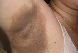 [Figure caption and citation for the preceding image starts]: Acanthosis nigricans involving the back and arms of an obese black womanFrom the collection of Miguel Gutierrez, MD [Citation ends].
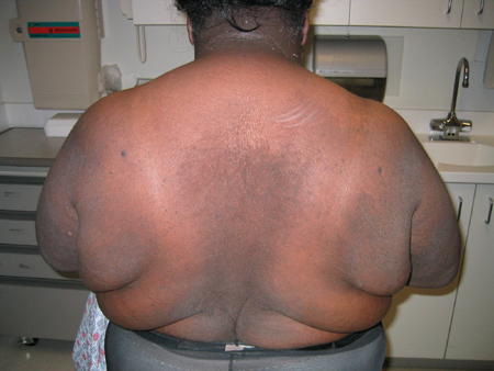
Malignancy screening
If no other cause is readily identifiable and there is rapid, widespread onset with involvement of mucosal surfaces and palms, malignancy screening for an intra-abdominal malignancy is warranted (e.g., abdominal CT scan following thorough clinical examination).[2]Schwartz RA. Acanthosis nigricans. J Am Acad Dermatol. 1994 Jul;31(1):1-19.
http://www.ncbi.nlm.nih.gov/pubmed/8021347?tool=bestpractice.com
[24]Schwartz RA. Acral acanthosis nigricans (acral acanthotic anomaly). J Am Acad Dermatol. 2007 Feb;56(2):349-50.
http://www.ncbi.nlm.nih.gov/pubmed/17224380?tool=bestpractice.com
Malignant AN may be accompanied by the sudden appearance of multiple seborrhoeic keratoses or generalised pruritus.[8]Moore RL, Devere TS. Epidermal manifestations of internal malignancy. Dermatol Clin. 2008 Jan;26(1):17-29.
http://www.ncbi.nlm.nih.gov/pubmed/18023768?tool=bestpractice.com
Patients with malignant AN may be underweight and are typically over the age of 40 years.[2]Schwartz RA. Acanthosis nigricans. J Am Acad Dermatol. 1994 Jul;31(1):1-19.
http://www.ncbi.nlm.nih.gov/pubmed/8021347?tool=bestpractice.com
[8]Moore RL, Devere TS. Epidermal manifestations of internal malignancy. Dermatol Clin. 2008 Jan;26(1):17-29.
http://www.ncbi.nlm.nih.gov/pubmed/18023768?tool=bestpractice.com
[9]Curth HO. Cancer associated with acanthosis nigricans: a review of the literature and report of a case of acanthosis nigricans with cancer of the breast. Arch Surg. 1943 Dec;47(6):517-52.
Testing for insulin resistance
Fasting glucose and insulin tests (e.g., fasting blood insulin) are performed if the patient has obesity to screen for insulin resistance.[18]Mukhtar Q, Cleverly G, Voorhees RE, et al. Prevalence of acanthosis nigricans and its association with hyperinsulinemia in New Mexico Adolescents. J Adolesc Health. 2001 May;28(5):372-6.
http://www.ncbi.nlm.nih.gov/pubmed/11336866?tool=bestpractice.com
Skin biopsy with histological examination
Skin biopsy is unnecessary in most cases, but can be helpful in atypical presentations or to rule out other differential diagnoses.[1]Das A, Datta D, Kassir M, et al. Acanthosis nigricans: a review. J Cosmet Dermatol. 2020 Aug;19(8):1857-65.
http://www.ncbi.nlm.nih.gov/pubmed/32516476?tool=bestpractice.com