Lichen sclerosus (LS) predominantly affects the genitals (but can also manifest as extragenital lesions).[1]Wallace HJ. Lichen sclerosus et atrophicus. Trans St Johns Hosp Dermatol Soc. 1971;57(1):9-30.
http://www.ncbi.nlm.nih.gov/pubmed/5570266?tool=bestpractice.com
[2]Powell JJ, Wojnarowska F. Lichen sclerosus. Lancet. 1999 May 22;353(9166):1777-83.
http://www.ncbi.nlm.nih.gov/pubmed/10348006?tool=bestpractice.com
Diagnosis of LS is typically clinical, but may be aided by skin biopsy if there is diagnostic uncertainty.
Always have a high index of suspicion for LS when a patient presents with skin lesions resembling white, crinkly/atrophic plaques, with or without associated symptoms of pruritus or pain; the patient may describe 'irritation'. Misdiagnosis/underdiagnosis is common.
Bear in mind that patients may present with extragenital skin lesions, but this is a less common manifestation of LS; around 6% to 15% of all cases are of isolated extragenital lesions.[1]Wallace HJ. Lichen sclerosus et atrophicus. Trans St Johns Hosp Dermatol Soc. 1971;57(1):9-30.
http://www.ncbi.nlm.nih.gov/pubmed/5570266?tool=bestpractice.com
[2]Powell JJ, Wojnarowska F. Lichen sclerosus. Lancet. 1999 May 22;353(9166):1777-83.
http://www.ncbi.nlm.nih.gov/pubmed/10348006?tool=bestpractice.com
Extragenital skin lesions are often asymptomatic but may be associated with pruritus. If a patient presents with extragenital disease, ask about anogenital symptoms as a part of the history and perform an examination of the genitalia in addition to a general skin examination.
Early diagnosis is key because initiating timely intervention leads to resolution of symptoms, as well as reduction or prevention of scarring, and reduces the risk of progression to malignancy in the majority of anogenital cases. Left untreated, anogenital LS can lead to scarring and obliteration of normal anatomical structures, such as loss of the labia minora and narrowing of the introitus in women, or phimosis in men; patients often first present with these features.
Occasionally, the first presentation of LS will be when squamous cell carcinoma (SCC) develops: a plaque, ulcer, or nodule arises and enlarges very quickly, sometimes within weeks. See Squamous cell carcinoma of the skin.
Consider key differential diagnoses, including vitiligo, lichen planus, and psoriasis; the condition may also mimic/be present alongside the autoimmune condition morphea. In men, also consider other causes of balanoposthitis, including other inflammatory skin conditions such as Zoon balanitis (plasma cell balanitis) or infections. See Balanoposthitis.
History
Take a comprehensive history; ask all patients with suspected LS about:
Other vulval/anogenital conditions/irritants, and anogenital cleaning methods
Many patients with LS will have other associated conditions (e.g., irritant contact dermatitis or allergic contact dermatitis), caused by self-management with non-prescription drugs and approaches.
Perfumed hygiene products and soaps may further exacerbate the condition and their use should also be enquired about.
Some patients may aggressively clean affected areas with a washcloth/sponge, causing further trauma to the skin.
Ask about symptoms of urinary or faecal incontinence, which may exacerbate the condition.[32]Bunker CB, Shim TN. Male genital lichen sclerosus. Indian J Dermatol. 2015 Mar-Apr;60(2):111-7.
http://www.ncbi.nlm.nih.gov/pubmed/25814697?tool=bestpractice.com
[33]Owen CM, Yell JA. Genital lichen sclerosus associated with incontinence. J Obstet Gynaecol. 2002 Mar;22(2):209-10.
http://www.ncbi.nlm.nih.gov/pubmed/12521711?tool=bestpractice.com
[34]van der Meijden WI, Boffa MJ, Ter Harmsel B, et al. 2021 European guideline for the management of vulval conditions. J Eur Acad Dermatol Venereol. 2022 Jul;36(7):952-72.
https://www.doi.org/10.1111/jdv.18102
http://www.ncbi.nlm.nih.gov/pubmed/35411963?tool=bestpractice.com
In some men, exposure to urine via urinary dribbling or incontinence has also been reported as a trigger for the condition.[34]van der Meijden WI, Boffa MJ, Ter Harmsel B, et al. 2021 European guideline for the management of vulval conditions. J Eur Acad Dermatol Venereol. 2022 Jul;36(7):952-72.
https://www.doi.org/10.1111/jdv.18102
http://www.ncbi.nlm.nih.gov/pubmed/35411963?tool=bestpractice.com
[35]Lewis FM, Tatnall FM, Velangi SS, et al. British Association of Dermatologists guidelines for the management of lichen sclerosus, 2018. Br J Dermatol. 2018 Apr;178(4):839-53.
https://www.doi.org/10.1111/bjd.16241
http://www.ncbi.nlm.nih.gov/pubmed/29313888?tool=bestpractice.com
Ask about other potential mechanical triggers/exacerbators, such as bicycle riding/wearing of tight clothing.[34]van der Meijden WI, Boffa MJ, Ter Harmsel B, et al. 2021 European guideline for the management of vulval conditions. J Eur Acad Dermatol Venereol. 2022 Jul;36(7):952-72.
https://www.doi.org/10.1111/jdv.18102
http://www.ncbi.nlm.nih.gov/pubmed/35411963?tool=bestpractice.com
A personal history of autoimmune conditions
LS in women has been associated with various autoimmune conditions and most commonly with autoimmune thyroid disorders.[23]Cooper SM, Ali I, Baldo M, et al. The association of lichen sclerosus and erosive lichen planus of the vulva with autoimmune disease: a case-control study. Arch Dermatol. 2008 Nov;144(11):1432-5.
https://www.doi.org/10.1001/archderm.144.11.1432
http://www.ncbi.nlm.nih.gov/pubmed/19015417?tool=bestpractice.com
A large case-control study of 765 women with LS found a statistically significant association of autoimmune conditions including autoimmune thyroiditis (odds ratio [OR] 2.67, P <0.001), hypothyroidism (OR 2.34, P <0.001), and hyperthyroidism (OR 2.05, P <0.001) compared with matched controls.[24]Fan R, Leasure AC, Maisha FI, et al. Thyroid disorders associated with lichen sclerosus: a case-control study in the All of Us Research Program. Br J Dermatol. 2022 Nov;187(5):797-9.
http://www.ncbi.nlm.nih.gov/pubmed/35661997?tool=bestpractice.com
LS may also mimic/be present alongside the autoimmune conditions morphea and vitiligo.
Autoimmune disorders may be more commonly found in women with LS compared with men with the condition.
One study of 532 patients found that LS was significantly more often associated with at least one autoimmune disease in women compared with men (OR 4.3, 95% CI 1.9 to 9.6).[25]Kreuter A, Kryvosheyeva Y, Terras S, et al. Association of autoimmune diseases with lichen sclerosus in 532 male and female patients. Acta Derm Venereol. 2013 Mar 27;93(2):238-41.
https://www.doi.org/10.2340/00015555-1512
http://www.ncbi.nlm.nih.gov/pubmed/23224274?tool=bestpractice.com
A history of metabolic syndrome
Obesity, and the associated condition metabolic syndrome, are more common in patients with LS.[15]Virgili A, Borghi A, Cazzaniga S, et al. New insights into potential risk factors and associations in genital lichen sclerosus: data from a multicentre Italian study on 729 consecutive cases. J Eur Acad Dermatol Venereol. 2017 Apr;31(4):699-704.
http://www.ncbi.nlm.nih.gov/pubmed/27515901?tool=bestpractice.com
[26]Hofer MD, Meeks JJ, Mehdiratta N, et al. Lichen sclerosus in men is associated with elevated body mass index, diabetes mellitus, coronary artery disease and smoking. World J Urol. 2014 Feb;32(1):105-8.
http://www.ncbi.nlm.nih.gov/pubmed/23633127?tool=bestpractice.com
[27]Ranum A, Freese R, Ramesh V, et al. Lichen sclerosus in female patients is associated with an increased risk of metabolic syndrome and cardiovascular comorbidities: a retrospective cohort review. Br J Dermatol. 2022 Dec;187(6):1030-2.
http://www.ncbi.nlm.nih.gov/pubmed/35947535?tool=bestpractice.com
One cross-sectional study found that adult patients with LS were more frequently overweight or obese (age-standardised prevalence ratio 1.44, 95% CI 1.30 to 1.59).[15]Virgili A, Borghi A, Cazzaniga S, et al. New insights into potential risk factors and associations in genital lichen sclerosus: data from a multicentre Italian study on 729 consecutive cases. J Eur Acad Dermatol Venereol. 2017 Apr;31(4):699-704.
http://www.ncbi.nlm.nih.gov/pubmed/27515901?tool=bestpractice.com
A family history of LS
Studies estimate that between 8% and 12% of women with LS have a first-degree relative with the condition.[19]Higgins CA, Cruickshank ME. A population-based case-control study of aetiological factors associated with vulval lichen sclerosus. J Obstet Gynaecol. 2012 Apr;32(3):271-5.
http://www.ncbi.nlm.nih.gov/pubmed/22369403?tool=bestpractice.com
[20]Sherman V, McPherson T, Baldo M, et al. The high rate of familial lichen sclerosus suggests a genetic contribution: an observational cohort study. J Eur Acad Dermatol Venereol. 2010 Sep;24(9):1031-4.
http://www.ncbi.nlm.nih.gov/pubmed/20202060?tool=bestpractice.com
Extragenital manifestations
Extragenital disease presents with atrophic white patches or plaques that are often asymptomatic, but sometimes may cause pruritus. Extragenital disease may involve any site on the body but is often found in flexural areas, the buttocks, thighs, breasts, trunk, and extremities.[11]Fistarol SK, Itin PH. Diagnosis and treatment of lichen sclerosus: an update. Am J Clin Dermatol. 2013 Feb;14(1):27-47.
https://www.doi.org/10.1007/s40257-012-0006-4
http://www.ncbi.nlm.nih.gov/pubmed/23329078?tool=bestpractice.com
[36]Pérez-López FR, Ceausu I, Depypere H, et al. EMAS clinical guide: vulvar lichen sclerosus in peri and postmenopausal women. Maturitas. 2013 Mar;74(3):279-82.
https://www.doi.org/10.1016/j.maturitas.2012.12.006
http://www.ncbi.nlm.nih.gov/pubmed/23291001?tool=bestpractice.com
LS may rarely affect the oral cavity, presenting as irregular whitish patches on the oral mucosa/lips.[7]Simms-Cendan J, Hoover K, Marathe K, et al. NASPAG clinical opinion: diagnosis and management of lichen sclerosis in pediatric and adolescent patients. J Pediatr Adolesc Gynecol. 2022 Apr;35(2):112-20.
http://www.ncbi.nlm.nih.gov/pubmed/34610442?tool=bestpractice.com
[37]Lagerstedt M, Karvinen K, Joki-Erkkilä M, et al. Childhood lichen sclerosus - a challenge for clinicians. Pediatr Dermatol. 2013 Jul-Aug;30(4):444-50.
http://www.ncbi.nlm.nih.gov/pubmed/23437870?tool=bestpractice.com
[38]Matela AM, Hagström J, Ruokonen H. Lichen sclerosus of the oral mucosa: clinical and histopathological findings. Review of the literature and a case report. Acta Odontol Scand. 2018 Jul;76(5):364-73.
http://www.ncbi.nlm.nih.gov/pubmed/29658796?tool=bestpractice.com
Approximately 20% of women with anogenital LS have extragenital lesions.[36]Pérez-López FR, Ceausu I, Depypere H, et al. EMAS clinical guide: vulvar lichen sclerosus in peri and postmenopausal women. Maturitas. 2013 Mar;74(3):279-82.
https://www.doi.org/10.1016/j.maturitas.2012.12.006
http://www.ncbi.nlm.nih.gov/pubmed/23291001?tool=bestpractice.com
Note that only around 6% to 15% of all cases are of isolated extragenital lesions.[1]Wallace HJ. Lichen sclerosus et atrophicus. Trans St Johns Hosp Dermatol Soc. 1971;57(1):9-30.
http://www.ncbi.nlm.nih.gov/pubmed/5570266?tool=bestpractice.com
[2]Powell JJ, Wojnarowska F. Lichen sclerosus. Lancet. 1999 May 22;353(9166):1777-83.
http://www.ncbi.nlm.nih.gov/pubmed/10348006?tool=bestpractice.com
LS may appear on areas of skin that have been exposed to trauma: for example, in operative scars or on skin exposed to tight clothing or other mechanical trauma.[11]Fistarol SK, Itin PH. Diagnosis and treatment of lichen sclerosus: an update. Am J Clin Dermatol. 2013 Feb;14(1):27-47.
https://www.doi.org/10.1007/s40257-012-0006-4
http://www.ncbi.nlm.nih.gov/pubmed/23329078?tool=bestpractice.com
[34]van der Meijden WI, Boffa MJ, Ter Harmsel B, et al. 2021 European guideline for the management of vulval conditions. J Eur Acad Dermatol Venereol. 2022 Jul;36(7):952-72.
https://www.doi.org/10.1111/jdv.18102
http://www.ncbi.nlm.nih.gov/pubmed/35411963?tool=bestpractice.com
The precipitation of disease activity in areas of trauma is known as the Koebner phenomenon.
Functional limitations and issues with mental health due to the condition
LS can have a significant impact on a patient’s quality of life and can lead to issues such as:[39]Fan R, Leasure AC, Maisha FI, et al. Depression and anxiety in patients with lichen sclerosus. JAMA Dermatol. 2022 Aug 1;158(8):953-4.
http://www.ncbi.nlm.nih.gov/pubmed/35704313?tool=bestpractice.com
[40]Vittrup G, Westmark S, Riis J, et al. The impact of psychosexual counseling in women lith lichen sclerosus: a randomized controlled trial. J Low Genit Tract Dis. 2022 Jul 1;26(3):258-64.
https://www.ncbi.nlm.nih.gov/pmc/articles/PMC9232275
http://www.ncbi.nlm.nih.gov/pubmed/35333024?tool=bestpractice.com
[41]Van de Nieuwenhof HP, Meeuwis KA, Nieboer TE, et al. The effect of vulvar lichen sclerosus on quality of life and sexual functioning. J Psychosom Obstet Gynaecol. 2010 Dec;31(4):279-84.
https://www.doi.org/10.3109/0167482X.2010.507890
http://www.ncbi.nlm.nih.gov/pubmed/20701461?tool=bestpractice.com
[42]Wijaya M, Lee G, Fischer G, et al. Quality of life in vulvar lichen sclerosus patients treated with long-term topical corticosteroids. J Low Genit Tract Dis. 2021 Apr 1;25(2):158-65.
http://www.ncbi.nlm.nih.gov/pubmed/33746196?tool=bestpractice.com
[43]Jabłonowska O, Woźniacka A, Szkarłat S, et al. Female genital lichen sclerosus is connected with a higher depression rate, decreased sexual quality of life and diminished work productivity. PLoS One. 2023;18(4):e0284948.
https://www.doi.org/10.1371/journal.pone.0284948
http://www.ncbi.nlm.nih.gov/pubmed/37098076?tool=bestpractice.com
[44]Arnold S, Fernando S, Rees S. Living with vulval lichen sclerosus: a qualitative interview study. Br J Dermatol. 2022 Dec;187(6):909-18.
https://www.doi.org/10.1111/bjd.21777
http://www.ncbi.nlm.nih.gov/pubmed/35831927?tool=bestpractice.com
Tailor further enquiry based on the sex and age of the patient as detailed below.
Adult women
Although most women with anogenital LS present with signs and symptoms of the condition, some may be asymptomatic (with the condition discovered incidentally).[45]Mauskar MM, Marathe K, Venkatesan A, et al. Vulvar diseases: conditions in adults and children. J Am Acad Dermatol. 2020 Jun;82(6):1287-98.
http://www.ncbi.nlm.nih.gov/pubmed/31712170?tool=bestpractice.com
Ask women about common anogenital symptoms, which may include:[3]Sheinis M, Selk A. Development of the Adult Vulvar Lichen Sclerosus Severity Scale - a Delphi consensus exercise for item generation. J Low Genit Tract Dis. 2018 Jan;22(1):66-73.
https://www.doi.org/10.1097/LGT.0000000000000361
http://www.ncbi.nlm.nih.gov/pubmed/29095746?tool=bestpractice.com
Pruritus (specifically ask if the patient feels the urge to scratch)
One cross-sectional study of 503 women aged between 18 and 50 years with biopsy-confirmed LS found that symptoms of pruritus prompted 31% to seek medical attention.[46]Krapf JM, Smith AB, Cigna ST, et al. Presenting symptoms and diagnosis of vulvar lichen sclerosus in premenopausal women: a cross-sectional study. J Low Genit Tract Dis. 2022 Jul 1;26(3):27175.
http://www.ncbi.nlm.nih.gov/pubmed/35533256?tool=bestpractice.com
Often worse at night and can disturb sleep.[35]Lewis FM, Tatnall FM, Velangi SS, et al. British Association of Dermatologists guidelines for the management of lichen sclerosus, 2018. Br J Dermatol. 2018 Apr;178(4):839-53.
https://www.doi.org/10.1111/bjd.16241
http://www.ncbi.nlm.nih.gov/pubmed/29313888?tool=bestpractice.com
Pain/burning/discomfort of the skin
This may cause difficulties with walking, sitting, and sexual intercourse, as well as causing issues such as anxiety and depression.[39]Fan R, Leasure AC, Maisha FI, et al. Depression and anxiety in patients with lichen sclerosus. JAMA Dermatol. 2022 Aug 1;158(8):953-4.
http://www.ncbi.nlm.nih.gov/pubmed/35704313?tool=bestpractice.com
[40]Vittrup G, Westmark S, Riis J, et al. The impact of psychosexual counseling in women lith lichen sclerosus: a randomized controlled trial. J Low Genit Tract Dis. 2022 Jul 1;26(3):258-64.
https://www.ncbi.nlm.nih.gov/pmc/articles/PMC9232275
http://www.ncbi.nlm.nih.gov/pubmed/35333024?tool=bestpractice.com
[41]Van de Nieuwenhof HP, Meeuwis KA, Nieboer TE, et al. The effect of vulvar lichen sclerosus on quality of life and sexual functioning. J Psychosom Obstet Gynaecol. 2010 Dec;31(4):279-84.
https://www.doi.org/10.3109/0167482X.2010.507890
http://www.ncbi.nlm.nih.gov/pubmed/20701461?tool=bestpractice.com
[42]Wijaya M, Lee G, Fischer G, et al. Quality of life in vulvar lichen sclerosus patients treated with long-term topical corticosteroids. J Low Genit Tract Dis. 2021 Apr 1;25(2):158-65.
http://www.ncbi.nlm.nih.gov/pubmed/33746196?tool=bestpractice.com
[43]Jabłonowska O, Woźniacka A, Szkarłat S, et al. Female genital lichen sclerosus is connected with a higher depression rate, decreased sexual quality of life and diminished work productivity. PLoS One. 2023;18(4):e0284948.
https://www.doi.org/10.1371/journal.pone.0284948
http://www.ncbi.nlm.nih.gov/pubmed/37098076?tool=bestpractice.com
[44]Arnold S, Fernando S, Rees S. Living with vulval lichen sclerosus: a qualitative interview study. Br J Dermatol. 2022 Dec;187(6):909-18.
https://www.doi.org/10.1111/bjd.21777
http://www.ncbi.nlm.nih.gov/pubmed/35831927?tool=bestpractice.com
Dysuria
Can occur in untreated disease due to urine coming into contact with inflamed and fissured/eroded skin.
In children, dysuria may lead to a fear of voiding and subsequent overflow incontinence.[7]Simms-Cendan J, Hoover K, Marathe K, et al. NASPAG clinical opinion: diagnosis and management of lichen sclerosis in pediatric and adolescent patients. J Pediatr Adolesc Gynecol. 2022 Apr;35(2):112-20.
http://www.ncbi.nlm.nih.gov/pubmed/34610442?tool=bestpractice.com
Dyspareunia (painful intercourse) with or without tearing and/or bleeding during intercourse
Can occur in sexually active women with anogenital LS at any time during the course of the condition; may be attributed to active skin inflammatory changes (causing atrophy, erosions, or fissures) or, although there is no robust evidence to support this, has been postulated to be due to resulting nerve and muscular changes in the setting of a chronic overlying inflammatory condition.[40]Vittrup G, Westmark S, Riis J, et al. The impact of psychosexual counseling in women lith lichen sclerosus: a randomized controlled trial. J Low Genit Tract Dis. 2022 Jul 1;26(3):258-64.
https://www.ncbi.nlm.nih.gov/pmc/articles/PMC9232275
http://www.ncbi.nlm.nih.gov/pubmed/35333024?tool=bestpractice.com
[47]Pope R, Lee MH, Myers A, et al. Lichen sclerosus and sexual dysfunction: a systematic review and meta-analysis. J Sex Med. 2022 Nov;19(11):1616-24.
http://www.ncbi.nlm.nih.gov/pubmed/36115787?tool=bestpractice.com
In one systematic review and meta-analysis, 59% of 486 women with LS reported sexual dysfunction.[47]Pope R, Lee MH, Myers A, et al. Lichen sclerosus and sexual dysfunction: a systematic review and meta-analysis. J Sex Med. 2022 Nov;19(11):1616-24.
http://www.ncbi.nlm.nih.gov/pubmed/36115787?tool=bestpractice.com
Dyspareunia can also develop due to functional limitations associated with scarring.[40]Vittrup G, Westmark S, Riis J, et al. The impact of psychosexual counseling in women lith lichen sclerosus: a randomized controlled trial. J Low Genit Tract Dis. 2022 Jul 1;26(3):258-64.
https://www.ncbi.nlm.nih.gov/pmc/articles/PMC9232275
http://www.ncbi.nlm.nih.gov/pubmed/35333024?tool=bestpractice.com
[46]Krapf JM, Smith AB, Cigna ST, et al. Presenting symptoms and diagnosis of vulvar lichen sclerosus in premenopausal women: a cross-sectional study. J Low Genit Tract Dis. 2022 Jul 1;26(3):27175.
http://www.ncbi.nlm.nih.gov/pubmed/35533256?tool=bestpractice.com
[47]Pope R, Lee MH, Myers A, et al. Lichen sclerosus and sexual dysfunction: a systematic review and meta-analysis. J Sex Med. 2022 Nov;19(11):1616-24.
http://www.ncbi.nlm.nih.gov/pubmed/36115787?tool=bestpractice.com
One cross-sectional study of 503 women with biopsy-confirmed LS found common presenting symptoms to include dyspareunia (68%) and tearing with intercourse or vaginal insertion (63%).[46]Krapf JM, Smith AB, Cigna ST, et al. Presenting symptoms and diagnosis of vulvar lichen sclerosus in premenopausal women: a cross-sectional study. J Low Genit Tract Dis. 2022 Jul 1;26(3):27175.
http://www.ncbi.nlm.nih.gov/pubmed/35533256?tool=bestpractice.com
Also ask about constipation, but bear in mind that adult women rarely present with symptoms of constipation and/or painful defecation.[36]Pérez-López FR, Ceausu I, Depypere H, et al. EMAS clinical guide: vulvar lichen sclerosus in peri and postmenopausal women. Maturitas. 2013 Mar;74(3):279-82.
https://www.doi.org/10.1016/j.maturitas.2012.12.006
http://www.ncbi.nlm.nih.gov/pubmed/23291001?tool=bestpractice.com
Constipation is more commonly seen in paediatric patients.
Adult men
In men, genital LS almost exclusively occurs in those who are uncircumcised (note that circumcision may be curative in male genital disease, particularly if carried out early).[31]Bunker CB. Diseases and disorders of the male genitalia. In: Fitzpatrick's dermatology in general medicine. New York, NY: McGraw-Hill; 2003: Chapter 3. [48]Riddell L, Edwards A, Sherrard J. Clinical features of lichen sclerosus in men attending a department of genitourinary medicine. Sex Transm Infect. 2000 Aug;76(4):311-3.
http://www.ncbi.nlm.nih.gov/pubmed/11026891?tool=bestpractice.com
LS of the penis may be asymptomatic, but diverse and vague symptomatology may be encountered.[48]Riddell L, Edwards A, Sherrard J. Clinical features of lichen sclerosus in men attending a department of genitourinary medicine. Sex Transm Infect. 2000 Aug;76(4):311-3.
http://www.ncbi.nlm.nih.gov/pubmed/11026891?tool=bestpractice.com
[49]Pelisse M. Lichen sclerosus [in French]. Ann Dermatol Venereol. 1987;114(3):411-9.
http://www.ncbi.nlm.nih.gov/pubmed/3605970?tool=bestpractice.com
[50]Edmonds EV, Hunt S, Hawkins D, et al. Clinical parameters in male genital lichen sclerosus: a case series of 329 patients. J Eur Acad Dermatol Venereol. 2012 Jun;26(6):730-7.
http://www.ncbi.nlm.nih.gov/pubmed/21707769?tool=bestpractice.com
LS in men (and boys) most commonly occurs on the glans penis, coronary sulcus, urethral meatus, and/or foreskin. Rarely, the penile shaft, perineal, scrotal, and perianal skin may be affected.[35]Lewis FM, Tatnall FM, Velangi SS, et al. British Association of Dermatologists guidelines for the management of lichen sclerosus, 2018. Br J Dermatol. 2018 Apr;178(4):839-53.
https://www.doi.org/10.1111/bjd.16241
http://www.ncbi.nlm.nih.gov/pubmed/29313888?tool=bestpractice.com
Pruritus is also relatively uncommon.[35]Lewis FM, Tatnall FM, Velangi SS, et al. British Association of Dermatologists guidelines for the management of lichen sclerosus, 2018. Br J Dermatol. 2018 Apr;178(4):839-53.
https://www.doi.org/10.1111/bjd.16241
http://www.ncbi.nlm.nih.gov/pubmed/29313888?tool=bestpractice.com
Men with untreated penile LS may develop sclerosis and narrowing of the foreskin, leading to phimosis or adhesions of the foreskin to the glans, which may cause sexual dysfunction/dyspareunia and urethral disease.[50]Edmonds EV, Hunt S, Hawkins D, et al. Clinical parameters in male genital lichen sclerosus: a case series of 329 patients. J Eur Acad Dermatol Venereol. 2012 Jun;26(6):730-7.
http://www.ncbi.nlm.nih.gov/pubmed/21707769?tool=bestpractice.com
Note that secondary phimosis in men is commonly due to LS.
Ask men about common symptoms of penile LS, including:[6]Shah M, van Bodegraven B. Male genital lichen sclerosus and associated symptoms range and severity: results of a questionnaire study. Skin Health Dis. 2023 Oct;3(5):e246.
https://www.doi.org/10.1002/ski2.246
http://www.ncbi.nlm.nih.gov/pubmed/37799358?tool=bestpractice.com
[35]Lewis FM, Tatnall FM, Velangi SS, et al. British Association of Dermatologists guidelines for the management of lichen sclerosus, 2018. Br J Dermatol. 2018 Apr;178(4):839-53.
https://www.doi.org/10.1111/bjd.16241
http://www.ncbi.nlm.nih.gov/pubmed/29313888?tool=bestpractice.com
Pain, dysuria, and/or changes in urinary stream, which may be caused by meatal stenosis (abnormal narrowing of the urethral opening)
Dyspareunia/sexual dysfunction
Colour changes in the affected area
Constrictive posthitis (foreskin tightening).
Enquire about a history of phimosis/paraphimosis.
Paediatric patients
Note that some paediatric patients (especially boys) may be asymptomatic, with lesions discovered incidentally by parents or carers.
In prepubertal girls, there may be a wide range of presenting complaints that may include the more classical signs and symptoms of skin changes and associated discomfort/irritation as described by adult women. In girls, ask about:[7]Simms-Cendan J, Hoover K, Marathe K, et al. NASPAG clinical opinion: diagnosis and management of lichen sclerosis in pediatric and adolescent patients. J Pediatr Adolesc Gynecol. 2022 Apr;35(2):112-20.
http://www.ncbi.nlm.nih.gov/pubmed/34610442?tool=bestpractice.com
Vulval discomfort/pain (may be worse at night)
Pruritus (may be worse at night)
Anogenital/vulval bleeding due to skin fissures
Constipation
Painful defecation (may lead to constipation)
Urinary tract symptoms such as dysuria, fear of voiding, and consequential overflow incontinence.
Like adult women, adolescent girls who are sexually active may also have symptoms of dyspareunia with associated lacerations at the base of the posterior fourchette due to trauma during intercourse.[7]Simms-Cendan J, Hoover K, Marathe K, et al. NASPAG clinical opinion: diagnosis and management of lichen sclerosis in pediatric and adolescent patients. J Pediatr Adolesc Gynecol. 2022 Apr;35(2):112-20.
http://www.ncbi.nlm.nih.gov/pubmed/34610442?tool=bestpractice.com
In boys with suspected LS, ask about phimosis. Bear in mind that a tight phimosis may hide typical skin changes described for adult men.[51]Dalziel K, Shaw S. Lichen sclerosus. BMJ. 2010 Feb 15;340:c731.
http://www.ncbi.nlm.nih.gov/pubmed/20156913?tool=bestpractice.com
Phimosis is the most frequent presentation of LS in boys, with reported incidence ranging from 12% to 100%.[9]Chalmers RJ, Burton PA, Bennett RF, et al. Lichen sclerosus et atrophicus. A common and distinctive cause of phimosis in boys. Arch Dermatol. 1984 Aug;120(8):1025-7.
http://www.ncbi.nlm.nih.gov/pubmed/6465907?tool=bestpractice.com
[35]Lewis FM, Tatnall FM, Velangi SS, et al. British Association of Dermatologists guidelines for the management of lichen sclerosus, 2018. Br J Dermatol. 2018 Apr;178(4):839-53.
https://www.doi.org/10.1111/bjd.16241
http://www.ncbi.nlm.nih.gov/pubmed/29313888?tool=bestpractice.com
[52]Kiss A, Király L, Kutasy B, et al. High incidence of balanitis xerotica obliterans in boys with phimosis: prospective 10-year study. Pediatr Dermatol. 2005 Jul-Aug;22(4):305-8.
http://www.ncbi.nlm.nih.gov/pubmed/16060864?tool=bestpractice.com
[53]Meuli M, Briner J, Hanimann B, et al. Lichen sclerosus et atrophicus causing phimosis in boys: a prospective study with 5-year followup after complete circumcision. J Urol. 1994 Sep;152(3):987-9.
http://www.ncbi.nlm.nih.gov/pubmed/8051779?tool=bestpractice.com
[54]Yardley IE, Cosgrove C, Lambert AW. Paediatric preputial pathology: are we circumcising enough? Ann R Coll Surg Engl. 2007 Jan;89(1):62-5.
https://www.ncbi.nlm.nih.gov/pmc/articles/PMC1963523
http://www.ncbi.nlm.nih.gov/pubmed/17316525?tool=bestpractice.com
Perianal involvement is rare.[35]Lewis FM, Tatnall FM, Velangi SS, et al. British Association of Dermatologists guidelines for the management of lichen sclerosus, 2018. Br J Dermatol. 2018 Apr;178(4):839-53.
https://www.doi.org/10.1111/bjd.16241
http://www.ncbi.nlm.nih.gov/pubmed/29313888?tool=bestpractice.com
Older boys who are sexually active may also present with symptoms of dyspareunia.
It is also important to note that sexual abuse may sometimes be suspected in children with LS presenting with ecchymoses (bleeding within the skin leading to bruising) and erosions, which may lead to a missed diagnosis of LS.[55]Powell J, Wojnarowska F. Childhood vulvar lichen sclerosus: an increasingly common problem. J Am Acad Dermatol. 2001 May;44(5):803-6.
http://www.ncbi.nlm.nih.gov/pubmed/11312428?tool=bestpractice.com
Be aware, however, that a diagnosis of LS does not exclude sexual abuse, and mechanical trauma due to sexual abuse has been implicated in both development and persistence of LS lesions secondary to the Koebner phenomenon.
Physical examination
Perform a thorough examination of all skin surfaces including genitalia and oral cavity in the patient with a suspected diagnosis of LS. LS predominantly affects the anogenital area, although there may be evidence of extragenital disease in a subset of patients.
Anogenital disease
Patients with anogenital LS will often present with both colour and texture change in the affected area.[3]Sheinis M, Selk A. Development of the Adult Vulvar Lichen Sclerosus Severity Scale - a Delphi consensus exercise for item generation. J Low Genit Tract Dis. 2018 Jan;22(1):66-73.
https://www.doi.org/10.1097/LGT.0000000000000361
http://www.ncbi.nlm.nih.gov/pubmed/29095746?tool=bestpractice.com
Signs vary depending on the degree of inflammation and the stage of disease/healing. These may include:[3]Sheinis M, Selk A. Development of the Adult Vulvar Lichen Sclerosus Severity Scale - a Delphi consensus exercise for item generation. J Low Genit Tract Dis. 2018 Jan;22(1):66-73.
https://www.doi.org/10.1097/LGT.0000000000000361
http://www.ncbi.nlm.nih.gov/pubmed/29095746?tool=bestpractice.com
[56]Kirtschig G, Kinberger M, Kreuter A, et al; European Dermatology Forum. EuroGuiDerm guideline on lichen sclerosus. Jun 2023 [internet publication].
https://www.guidelines.edf.one//uploads/attachments/clmub3q4l0an5uhjrluc4r0yq-lichen-sclerosus-gl.pdf
Purpura/ecchymoses
Pathognomonic of the condition; commonly due to scratching-related trauma secondary to pruritus.
May develop due to trauma to affected skin during sexual intercourse or defecation.[57]Nair PA. Vulvar Lichen sclerosus et atrophicus. J Midlife Health. 2017 Apr-Jun;8(2):55-62.
https://www.doi.org/10.4103/jmh.JMH_13_17
http://www.ncbi.nlm.nih.gov/pubmed/28706405?tool=bestpractice.com
Whitening of skin
May be mistaken for vitiligo.
In some women, patches of whitening may become confluent and extend around the vulval/perianal skin, with a 'figure of eight' appearance.
Changes in skin texture such as atrophy, sclerosis, lichenification, and/or hyperkeratosis
Crinkly/fine wrinkling of skin is classical, but there may be smooth, shiny/waxy, or hyperkeratotic changes.
Excoriations
Fissures (linear breaks in the skin)
Erosions (circumscribed lesions due to intraepithelial loss of the epidermis; usually depressed)
Ulcerations (shallow open areas of skin due to full thickness loss of the epidermis and damage to the dermis)
Erythema (redness of skin)
As the condition progresses, scarring of the anogenital area may also occur in women and girls with untreated LS.[3]Sheinis M, Selk A. Development of the Adult Vulvar Lichen Sclerosus Severity Scale - a Delphi consensus exercise for item generation. J Low Genit Tract Dis. 2018 Jan;22(1):66-73.
https://www.doi.org/10.1097/LGT.0000000000000361
http://www.ncbi.nlm.nih.gov/pubmed/29095746?tool=bestpractice.com
Look for such changes, including:
Clitoral hood fusion
Labia fusion/resorption
Narrowing of the introitus
Anterior changes (may include the clitoral hood, often extending to midline agglutination of labia majora between clitoral unit and vestibule anterior to urethra; LS does not involve the clitoris)
Perianal involvement
Formation of posterior commissure bands/fourchette webs.
Scarring and subsequent loss of normal anatomical structure may be seen in untreated disease and usually develops in the setting of chronic inflammation, often with silent progression.[58]Cooper SM, Gao XH, Powell JJ, et al. Does treatment of vulvar lichen sclerosus influence its prognosis? Arch Dermatol. 2004 Jun;140(6):702-6.
https://www.doi.org/10.1001/archderm.140.6.702
http://www.ncbi.nlm.nih.gov/pubmed/15210461?tool=bestpractice.com
Once scarring develops, it cannot be reversed.
[Figure caption and citation for the preceding image starts]: Vulval lichen sclerosus showing white plaques and scarring with some loss of the labia minoraDalziel K et al. BMJ 2010; 340: c731; used with permission [Citation ends].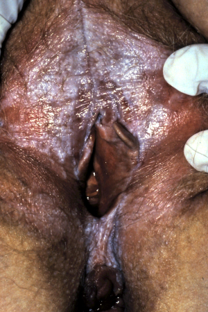
In men/boys, LS may cause constrictive posthitis (foreskin tightening) leading to paraphimosis or phimosis.[8]Celis S, Reed F, Murphy F, et al. Balanitis xerotica obliterans in children and adolescents: a literature review and clinical series. J Pediatr Urol. 2014 Feb;10(1):34-9.
https://www.doi.org/10.1016/j.jpurol.2013.09.027
http://www.ncbi.nlm.nih.gov/pubmed/24295833?tool=bestpractice.com
[9]Chalmers RJ, Burton PA, Bennett RF, et al. Lichen sclerosus et atrophicus. A common and distinctive cause of phimosis in boys. Arch Dermatol. 1984 Aug;120(8):1025-7.
http://www.ncbi.nlm.nih.gov/pubmed/6465907?tool=bestpractice.com
[Figure caption and citation for the preceding image starts]: Lichen sclerosus of glans penis in adult showing whiteness and purpuraDalziel K et al. BMJ 2010; 340: c731; used with permission [Citation ends].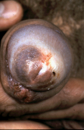
Extragenital disease
Fully inspect all areas of the skin for extragenital disease.
Extragenital plaques are typically depigmented but may also present with hyperkeratosis and follicular plugging.
Extragenital disease may affect any site on the body; it is most typically seen in flexural areas, the buttocks, thighs, breasts, trunk, and extremities.[11]Fistarol SK, Itin PH. Diagnosis and treatment of lichen sclerosus: an update. Am J Clin Dermatol. 2013 Feb;14(1):27-47.
https://www.doi.org/10.1007/s40257-012-0006-4
http://www.ncbi.nlm.nih.gov/pubmed/23329078?tool=bestpractice.com
A specialist may use dermoscopy in patients with suspected LS; dermoscopy may reveal structureless white to yellow areas (most commonly), chrysalis-like structures, linear irregular vessels, and perifollicular scaling.[59]Mahajan SA, Dave JS. Dermoscopic evaluation of extragenital lichen sclerosus et atrophicus. Dermatol Pract Concept. 2022 Jul;12(3):e2022125.
https://www.doi.org/10.5826/dpc.1203a125
http://www.ncbi.nlm.nih.gov/pubmed/36159134?tool=bestpractice.com
[60]Garrido-Ríos AA, Alvarez-Garrido H, Sanz-Muñoz C, et al. Dermoscopy of extragenital lichen sclerosus. Arch Dermatol. 2009 Dec;145(12):1468.
http://www.ncbi.nlm.nih.gov/pubmed/20026867?tool=bestpractice.com
Examine the lips and oral cavity; look for irregular whitish patches on the oral mucosa/lips.
Rarely, LS may affect the oral cavity.[7]Simms-Cendan J, Hoover K, Marathe K, et al. NASPAG clinical opinion: diagnosis and management of lichen sclerosis in pediatric and adolescent patients. J Pediatr Adolesc Gynecol. 2022 Apr;35(2):112-20.
http://www.ncbi.nlm.nih.gov/pubmed/34610442?tool=bestpractice.com
[37]Lagerstedt M, Karvinen K, Joki-Erkkilä M, et al. Childhood lichen sclerosus - a challenge for clinicians. Pediatr Dermatol. 2013 Jul-Aug;30(4):444-50.
http://www.ncbi.nlm.nih.gov/pubmed/23437870?tool=bestpractice.com
[38]Matela AM, Hagström J, Ruokonen H. Lichen sclerosus of the oral mucosa: clinical and histopathological findings. Review of the literature and a case report. Acta Odontol Scand. 2018 Jul;76(5):364-73.
http://www.ncbi.nlm.nih.gov/pubmed/29658796?tool=bestpractice.com
Look for signs of Koebnerisation (the precipitation of disease activity in areas of trauma).
LS may appear on skin subjected to trauma: for example, in operative scars, on areas of skin exposed to tight clothing, or from bicycle saddles in cyclists, or other mechanical trauma.[11]Fistarol SK, Itin PH. Diagnosis and treatment of lichen sclerosus: an update. Am J Clin Dermatol. 2013 Feb;14(1):27-47.
https://www.doi.org/10.1007/s40257-012-0006-4
http://www.ncbi.nlm.nih.gov/pubmed/23329078?tool=bestpractice.com
[34]van der Meijden WI, Boffa MJ, Ter Harmsel B, et al. 2021 European guideline for the management of vulval conditions. J Eur Acad Dermatol Venereol. 2022 Jul;36(7):952-72.
https://www.doi.org/10.1111/jdv.18102
http://www.ncbi.nlm.nih.gov/pubmed/35411963?tool=bestpractice.com
Initial investigations
Diagnosis is usually based on clinical findings. Have a high index of suspicion when patients present with depigmented crinkly texture changes to the skin (in the anogenital areas or less commonly in extragenital locations) or with architectural changes in the anogenital area.[45]Mauskar MM, Marathe K, Venkatesan A, et al. Vulvar diseases: conditions in adults and children. J Am Acad Dermatol. 2020 Jun;82(6):1287-98.
http://www.ncbi.nlm.nih.gov/pubmed/31712170?tool=bestpractice.com
Indications for biopsy include:[35]Lewis FM, Tatnall FM, Velangi SS, et al. British Association of Dermatologists guidelines for the management of lichen sclerosus, 2018. Br J Dermatol. 2018 Apr;178(4):839-53.
https://www.doi.org/10.1111/bjd.16241
http://www.ncbi.nlm.nih.gov/pubmed/29313888?tool=bestpractice.com
[56]Kirtschig G, Kinberger M, Kreuter A, et al; European Dermatology Forum. EuroGuiDerm guideline on lichen sclerosus. Jun 2023 [internet publication].
https://www.guidelines.edf.one//uploads/attachments/clmub3q4l0an5uhjrluc4r0yq-lichen-sclerosus-gl.pdf
[61]American College of Obstericians and Gynecologists. Diagnosis and management of vulvar skin disorders: ACOG practice bulletin summary, number 224. Obstet Gynecol. 2020 Jul;136(1):222-5.
http://www.ncbi.nlm.nih.gov/pubmed/32590722?tool=bestpractice.com
Immunohistochemistry with CD34 following biopsy may be useful for differentiating LS from other lichenoid dermatitides such as lichen planus, as CD34 expression is often absent in areas of affected visible collagen.[62]Borretta LJ, Crawford RI. CD34 Staining in disorders of collagen degeneration other than morphea. Am J Dermatopathol. 2020 Aug;42(8):623-4.
https://www.doi.org/10.1097/DAD.0000000000001584
http://www.ncbi.nlm.nih.gov/pubmed/32701705?tool=bestpractice.com
[63]Weyers W. Hypertrophic lichen sclerosus sine sclerosis: clues to histopathologic diagnosis when presenting as psoriasiform lichenoid dermatitis. J Cutan Pathol. 2015 Feb;42(2):118-29.
http://www.ncbi.nlm.nih.gov/pubmed/25413733?tool=bestpractice.com
Note that biopsy samples are generally not taken from children with suspected LS; however, refer children who do not respond to initial therapy or in whom there is diagnostic doubt for specialist input and consideration of biopsy (i.e., to a paediatric dermatologist or paediatric gynaecologist with expertise in vulval disorders).[7]Simms-Cendan J, Hoover K, Marathe K, et al. NASPAG clinical opinion: diagnosis and management of lichen sclerosis in pediatric and adolescent patients. J Pediatr Adolesc Gynecol. 2022 Apr;35(2):112-20.
http://www.ncbi.nlm.nih.gov/pubmed/34610442?tool=bestpractice.com
Specialist referral
If biopsy is indicated, the results will usually confirm the diagnosis. However, if biopsy does not provide definitive diagnosis, refer the patient to a specialist, who may consider further investigations.
If the diagnosis remains unclear following further investigations, then clinical judgement should determine next steps. Some experts advocate a trial of treatment to help diagnose the condition in these instances, given most cases of LS are responsive to topical corticosteroids. However, this is not a widely accepted approach because there are other conditions, including some differentials, which also respond to topical corticosteroids.
Emerging tests
Emerging diagnostic methods that are not routinely used at present but have shown promise in small preliminary studies include non-invasive imaging techniques such as optical coherence tomography (OCT) and high-frequency ultrasound.[64]Huisman BW, Pagan L, Naafs RGC, et al. Dermatoscopy and optical coherence tomography in vulvar high-grade squamous intraepithelial lesions and lichen sclerosus: a prospective observational trial. J Low Genit Tract Dis. 2023 Jul 1;27(3):255-61.
https://www.doi.org/10.1097/LGT.0000000000000731
http://www.ncbi.nlm.nih.gov/pubmed/36924426?tool=bestpractice.com
[65]Huisman BW, Pagan L, Ulrich M, et al. Reflectance confocal microscopy as a non-invasive imaging tool in vulvar high-grade squamous intraepithelial lesions and lichen sclerosus: a descriptive morphological study in patients and healthy volunteers. Exp Dermatol. 2023 Oct;32(10):1734-43.
https://www.doi.org/10.1111/exd.14888
http://www.ncbi.nlm.nih.gov/pubmed/37486173?tool=bestpractice.com
[66]Zhou MY, Wang YK, Zhu QL, et al. High-frequency ultrasound features in vulvar lichen sclerosus and correlation with histopathology. Skin Res Technol. 2022 Nov;28(6):780-5.
https://www.doi.org/10.1111/srt.13198
http://www.ncbi.nlm.nih.gov/pubmed/35969183?tool=bestpractice.com
One small observational study that included 25 women, 10 of whom had LS, used a combination of OCT and dermatoscopy to define characteristics to aid differentiation of affected skin from healthy vulvar skin, which could complement clinical assessment.[64]Huisman BW, Pagan L, Naafs RGC, et al. Dermatoscopy and optical coherence tomography in vulvar high-grade squamous intraepithelial lesions and lichen sclerosus: a prospective observational trial. J Low Genit Tract Dis. 2023 Jul 1;27(3):255-61.
https://www.doi.org/10.1097/LGT.0000000000000731
http://www.ncbi.nlm.nih.gov/pubmed/36924426?tool=bestpractice.com
One high-frequency ultrasound study of lesions from 40 patients with confirmed LS found that a hypoechoic dermal band was present in all lesions, with a significant linear positive correlation between the histopathological depth and corresponding hypoechoic dermal band thickness. The authors concluded that high-frequency ultrasound characteristics may provide valuable information in the precise diagnosis and the treatment monitoring of vulval LS.[66]Zhou MY, Wang YK, Zhu QL, et al. High-frequency ultrasound features in vulvar lichen sclerosus and correlation with histopathology. Skin Res Technol. 2022 Nov;28(6):780-5.
https://www.doi.org/10.1111/srt.13198
http://www.ncbi.nlm.nih.gov/pubmed/35969183?tool=bestpractice.com
Further assessment of the utility and applicabilities of these diagnostic methods is warranted.
Pitfalls
Watch for patients presenting without classical signs and symptoms or histological features of LS.
In particular, note that some patients with LS may have non-specific erythema and linear fissures (mimicking other skin conditions such as dermatitis) without classic late-stage scarring, but present with dyspareunia; these patients often have negative biopsy results.[67]Day T, Selim MA, Allbritton JI, et al. Nonsclerotic lichen sclerosus: definition of a concept and pathologic description. J Low Genit Tract Dis. 2023 Oct 1;27(4):358-64.
https://www.doi.org/10.1097/LGT.0000000000000760
http://www.ncbi.nlm.nih.gov/pubmed/37467474?tool=bestpractice.com
Some patients may have asymptomatic skin lesions.[45]Mauskar MM, Marathe K, Venkatesan A, et al. Vulvar diseases: conditions in adults and children. J Am Acad Dermatol. 2020 Jun;82(6):1287-98.
http://www.ncbi.nlm.nih.gov/pubmed/31712170?tool=bestpractice.com
Be aware that there may be significant delay between disease onset and presentation, owing to patient embarrassment or in some cases due to the clinician’s lack of familiarity with the condition/failure to examine the genital skin.[15]Virgili A, Borghi A, Cazzaniga S, et al. New insights into potential risk factors and associations in genital lichen sclerosus: data from a multicentre Italian study on 729 consecutive cases. J Eur Acad Dermatol Venereol. 2017 Apr;31(4):699-704.
http://www.ncbi.nlm.nih.gov/pubmed/27515901?tool=bestpractice.com
Patients with untreated disease may present with scarring or architectural changes such as clitoral hood fusion, labia fusion/resorption, narrowing of the introitus, anterior changes (may include the clitoral hood, often extending to midline agglutination of labia majora between clitoral unit and vestibule anterior to urethra; note that LS never affects the clitoris itself), and formation of posterior commissure bands.[3]Sheinis M, Selk A. Development of the Adult Vulvar Lichen Sclerosus Severity Scale - a Delphi consensus exercise for item generation. J Low Genit Tract Dis. 2018 Jan;22(1):66-73.
https://www.doi.org/10.1097/LGT.0000000000000361
http://www.ncbi.nlm.nih.gov/pubmed/29095746?tool=bestpractice.com
[35]Lewis FM, Tatnall FM, Velangi SS, et al. British Association of Dermatologists guidelines for the management of lichen sclerosus, 2018. Br J Dermatol. 2018 Apr;178(4):839-53.
https://www.doi.org/10.1111/bjd.16241
http://www.ncbi.nlm.nih.gov/pubmed/29313888?tool=bestpractice.com
Bear in mind that LS is frequently misdiagnosed; in particular:
Patients with LS who have non-specific clinical signs, such as linear fissures and erythema, may be misdiagnosed with vulvovaginal yeast infection (candidiasis).[46]Krapf JM, Smith AB, Cigna ST, et al. Presenting symptoms and diagnosis of vulvar lichen sclerosus in premenopausal women: a cross-sectional study. J Low Genit Tract Dis. 2022 Jul 1;26(3):27175.
http://www.ncbi.nlm.nih.gov/pubmed/35533256?tool=bestpractice.com
Consider early LS in patients with suspected fungal infection who have a negative potassium hydroxide (KOH) preparation or culture but continue to have symptoms such as pruritus or soreness.
One cross-sectional study of 503 women with biopsy-confirmed LS found, on average, a 4-year delay between symptom onset and diagnosis, with 66% initially receiving an alternative diagnosis; 49% of these women were misdiagnosed as having a vulvovaginal yeast infection.[46]Krapf JM, Smith AB, Cigna ST, et al. Presenting symptoms and diagnosis of vulvar lichen sclerosus in premenopausal women: a cross-sectional study. J Low Genit Tract Dis. 2022 Jul 1;26(3):27175.
http://www.ncbi.nlm.nih.gov/pubmed/35533256?tool=bestpractice.com
LS occurring in extragenital sites alongside morphea can be misdiagnosed as morphea alone (also known as localised scleroderma), which has a similar clinical and histological presentation to LS.[68]Khan Mohammad Beigi P. The immunogenetics of morphea and lichen sclerosus. Adv Exp Med Biol. 2022;1367:155-72.
http://www.ncbi.nlm.nih.gov/pubmed/35286696?tool=bestpractice.com
[69]Lutz V, Francès C, Bessis D, et al. High frequency of genital lichen sclerosus in a prospective series of 76 patients with morphea: toward a better understanding of the spectrum of morphea. Arch Dermatol. 2012 Jan;148(1):24-8.
https://www.doi.org/10.1001/archdermatol.2011.305
http://www.ncbi.nlm.nih.gov/pubmed/22004877?tool=bestpractice.com
In one study of 76 patients diagnosed with morphea, 45% were also found to have previously undiagnosed LS.[69]Lutz V, Francès C, Bessis D, et al. High frequency of genital lichen sclerosus in a prospective series of 76 patients with morphea: toward a better understanding of the spectrum of morphea. Arch Dermatol. 2012 Jan;148(1):24-8.
https://www.doi.org/10.1001/archdermatol.2011.305
http://www.ncbi.nlm.nih.gov/pubmed/22004877?tool=bestpractice.com
Irritation, dryness, pruritus, and dyspareunia may occur as a part of the genitourinary syndrome of menopause (GSM) and misdiagnosis of LS as GSM (and vice versa) may occur when these symptoms and associated signs are present.[42]Wijaya M, Lee G, Fischer G, et al. Quality of life in vulvar lichen sclerosus patients treated with long-term topical corticosteroids. J Low Genit Tract Dis. 2021 Apr 1;25(2):158-65.
http://www.ncbi.nlm.nih.gov/pubmed/33746196?tool=bestpractice.com
Numerous other dermatological conditions affecting the anogenital area may present in a similar fashion to LS - see Differentials.