Criteria
Histological criteria[6]
Diagnosis is confirmed by the presence of the following characteristic pathohistological findings, which identify three histological subtypes of microscopic colitis:[6]
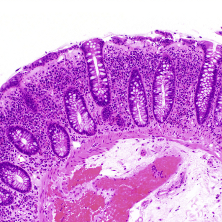
Collagenous colitis: characterised by a thickened subepithelial collagenous band of ≥10 micrometres (normal <5 micrometres) combined with an increased inflammatory infiltrate in the lamina propria.
[Figure caption and citation for the preceding image starts]: Biopsy demonstrating collagenous colitis with a thickened subepithelial collagen bandTome J et al. Microscopic Colitis: A Concise Review for Clinicians. Mayo Clin Proc. 2021 May;96(5):1302-8; used with permission [Citation ends].
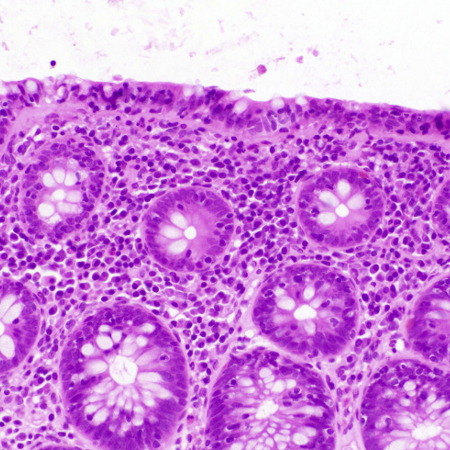
Lymphocytic colitis: characterised by an increased number of intraepithelial lymphocytes of ≥20 per 100 surface epithelial cells (normal <5 micrometres) combined with an increased inflammatory infiltrate in the lamina propria. The collagenous band is not significantly thickened (<10 micrometres).
[Figure caption and citation for the preceding image starts]: Biopsy demonstrating lymphocytic colitis with intraepithelial lymphocytosisTome J et al. Microscopic Colitis: A Concise Review for Clinicians. Mayo Clin Proc. 2021 May;96(5):1302-8; used with permission [Citation ends].
Microscopic colitis incomplete (also known as microscopic colitis not otherwise specified): comprises incomplete collagenous colitis (defined by a thickened subepithelial collagenous band of >5 micrometres but <10 micrometres) and incomplete lymphocytic colitis (defined by >10 but <20 intraepithelial lymphocytes per 100 epithelial cells and a normal collagenous band). Both types show a mild inflammatory infiltrate in the lamina propria.
Use of this content is subject to our disclaimer