Recommendations
Key Recommendations
Consider acute aspiration in any patient with sudden onset of fever, wheezing, and/or cyanosis, or radiological changes of consolidation, who has risk factors for aspiration (e.g., swallowing dysfunction, impaired conscious level, substance misuse). However, be aware that many patients are asymptomatic following aspiration.[2]
Have a high index of suspicion for acute aspiration and take prompt action if this is witnessed or suspected, particularly if the patient has a reduced conscious level.
Take a focused history to make the diagnosis of acute aspiration. However, this should be corroborated using imaging.
Order a chest x-ray first-line, but consider chest computed tomography if the patient fails to improve with initial treatment, to rule out complications such as empyema or lung abscess.
Organise early bronchoscopy if you suspect the patient has aspirated barium during gastrointestinal studies.
If the patient has aspirated a foreign body, see our topic Foreign body aspiration.
Note that, although some patients may have significant signs and symptoms following aspiration, many are asymptomatic.[2] Have a high index of suspicion for acute aspiration and take prompt action if this is witnessed or suspected, particularly if the patient has a reduced conscious level.
Consider acute aspiration in any patient with sudden onset of fever, wheezing, and/or cyanosis who has risk factors for aspiration. Note that symptoms and signs may be transient. Risk factors for aspiration can be classified according to the underlying mechanism, which can cause acute aspiration due to:
Aspiration of food, drink, oral secretions, or other substances:
Swallowing dysfunction (e.g., stroke, dementia, epilepsy, multiple sclerosis, Parkinson’s disease, motor neuron disease, cardiorespiratory disease, head and neck cancers)
Impaired conscious level
Substance misuse (e.g., acute alcohol intoxication, opioid toxicity)
During general anaesthesia (or other oropharyngeal procedures) or in the intensive care unit. These are common scenarios; the patient may present asymptomatically, or with bronchospasm, hypoxia, cough, dyspnoea, fever, and even respiratory failure from non-cardiogenic pulmonary oedema[11][12]
Gastrointestinal (GI) disease (e.g., upper GI surgery, hiatal hernia), conditions that affect gastric emptying (e.g., obesity, pregnancy), and oesophageal abnormalities (e.g., dysmotility, strictures, fistulas, gastroparesis [which can be due to diabetes])
Poor cough (e.g., upper airway problems such as pharyngeal pouches and vocal cord palsies, neuromuscular disease)
Increased severity of illness
Following upper gastrointestinal studies with barium. Aspiration of barium sulfate can cause aspiration pneumonitis, which can present with respiratory distress
Head and neck trauma. The patient may acutely aspirate blood in this scenario
Reflux/vomiting:
Substance misuse (e.g., acute alcohol intoxication, opioid toxicity)
General anaesthetic or oropharyngeal procedures
Recumbent position during enteral feeding
Polytrauma
Head and neck cancers.
Other features of acute aspiration include intractable cough, dyspnoea, crackles heard on auscultation of the lungs, reduced conscious level, and acute respiratory distress syndrome (ARDS).
Practical tip
A focused history is key to the diagnosis of acute aspiration because the signs, symptoms, and results of imaging can be indistinguishable from other causes of ARDS.
Monitor the patient for the development of aspiration pneumonia following acute aspiration, but note that not all episodes of aspiration lead to an infection. Always consider acute aspiration as a cause of pneumonia in a patient who is older with comorbidities. See our topic Aspiration pneumonia.
[Figure caption and citation for the preceding image starts]: Bronchoscopy showing barium aspiration in a lung transplant patient in the right mainstem bronchus after a barium swallow studyFrom the collection of Dr Kamran Mahmood [Citation ends].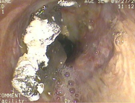
Chest x-ray
Take a focused history to make the diagnosis of acute aspiration. However, this should be corroborated using imaging; order a chest x-ray as first-line.
Be aware that chest x-ray changes may only be seen if acute aspiration has led to aspiration pneumonitis or pneumonia. Bear in mind that aspiration pneumonitis and pneumonia can also co-exist.
Also note that the pattern of chest x-ray changes varies depending on the positioning of the patient when they aspirate, and the mechanism of aspiration.
Look for patchy, bilateral airspace consolidations with a perihilar and basilar distribution following aspiration.[27]
In general, the right lung is involved more frequently than the left lung. Any material that is aspirated is more likely to end up in the right lung because:
The angle between the trachea and the right main bronchus is obtuse
The right main bronchus is wider and more vertical than the left main bronchus.
The most commonly involved segments are the superior and posterobasal segments of the right lower lobe and the posterior segment of the right upper lobe, because of their dependent location if the patient aspirates while lying in a supine position. However, other segments can be involved if the patient is not lying in a supine position. For example, if the patient is lying on the left side, the left lung may show consolidation.
In acute on chronic aspiration, the consolidation may predominantly be in the basal segments.
Barium aspiration may show dense opacities in the mid- and lower lungs, with a miliary pattern.[38][39]
[Figure caption and citation for the preceding image starts]: A. Portable upright chest x-ray before aspiration; B. Chest x-ray 1 hour after aspiration, showing bilateral diffuse alveolar infiltrates, worse at the bases on the right sideFrom the collection of Dr Henri Colt [Citation ends].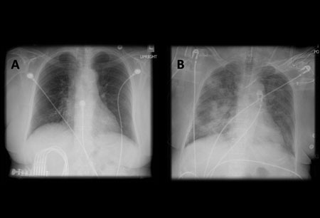
Chest x-ray findings of aspiration pneumonitis usually develop within 2 hours of aspiration and sometimes resolve quickly. However, opacities due to aspiration pneumonia can develop days later and can take weeks to resolve.
Blood tests
Order blood tests to differentiate between chemical pneumonitis and pneumonia; high white cell count, procalcitonin, or C-reactive protein indicates infection.
Chest computed tomography (CT)
Order chest CT if the patient does not improve with initial treatment, to rule out complications of aspiration such as empyema or lung abscess. Chest CT is better than chest-ray for visualising these complications.
Chest CT may show:
Extensive consolidation with cavitation if a lung abscess is present
Pleural effusion, often with loculations, if an empyema is present
Opacities in the posterior segments of the upper lobes and the superior segments of the lower lobes if a chemical pneumonitis or pneumonia is present. CT can precisely delineate the location of the lobar and segmental opacities. If the patient has significant aspiration, CT findings may be indistinguishable from those of acute respiratory distress syndrome (ARDS). Note that the pattern of changes on CT varies depending on the positioning of the patient when they aspirate and the mechanism of aspiration - see Chest x-ray above
Thickened interlobular septae, subpleural lines, and subpleural cysts following aspiration of barium. These findings may be present for a long time (even 1 year) after aspiration
Aspiration of fat or contrast material, which can sometimes be determined by measuring the tissue attenuation.[40]
Bronchoscopy
If you suspect a substantial amount of gastric content (>20-25 mL) is likely to have been aspirated, organise urgent (within a few hours) bronchoscopy and suctioning to remove any aspirated gastric fluid and solid material from the central airways.
Organise early bronchoscopy if you suspect the patient has aspirated barium during gastrointestinal studies, to remove the barium from their airway.
Get help from the critical care team if the patient has significant hypoxia or respiratory distress because bronchoscopy may cause further deterioration.
Other indications for bronchoscopy are to:
Clear the airway if the aspirated material is particulate or if there is radiographic evidence of lobar or segmental collapse
Collect quantitative cultures on bronchoalveolar lavage or protected specimen brush, which can be used to guide antibiotic therapy, particularly in patients who do not respond to empirical antibiotic treatment
Investigate alternative diagnoses that can cause a similar radiographic pattern to acute aspiration. These include ARDS imitators such as COVID-19 pneumonitis/pneumonia, acute interstitial pneumonitis (Hamman-Rich syndrome), acute eosinophilic pneumonia, cryptogenic organising pneumonia, diffuse alveolar haemorrhage, and acute hypersensitivity pneumonitis.[41]
[Figure caption and citation for the preceding image starts]: Bronchoscopy showing barium aspiration in a lung transplant patient in the right mainstem bronchus after a barium swallow studyFrom the collection of Dr Kamran Mahmood [Citation ends].
Arterial blood gas
May show hypoxaemia.
Use of this content is subject to our disclaimer