Recommendations
Key Recommendations
Suspect atrial flutter if the patient has:[5]
Palpitations
Dypnoea
Fatigue
Light-headedness
Chest discomfort
Altered consciousness
Polyuria.
Other features include:
Exercise intolerance
Worsening symptoms of heart failure
Features of an embolic event (such as stroke or pulmonary embolism – see our topics Ischaemic stroke and Pulmonary embolism).
Perform a 12-lead resting ECG for any patient with suspected atrial flutter.[5]
This will show continuous regular electrical activity (if a regular atrioventricular [AV] block is present), most commonly a saw-tooth pattern.[5]
Typically there is 2:1 AV block and the characteristic ventricular rate is 150 bpm. However, variable block may occur leading to an irregular rate.
The ECG pattern may fluctuate between atrial flutter and atrial fibrillation. See our topic New-onset atrial fibrillation.
Other classic ECG signs depend on the type of flutter.[5]
Urgently assess for features that suggest haemodynamic instability:[5][20]
Shock – see our topic Shock
Syncope
Myocardial ischaemia – see our topics ST-elevation myocardial infarction and Non ST-elevation myocardial infarction
Acute, severe heart failure (characterised by acute pulmonary oedema or raised jugular venous pressure) – see our topic Acute heart failure.
Patients with features of haemodynamic instability may need emergency electrical cardioversion.[5] See Haemodynamically unstable under Management recommendations. Seek advice from a cardiology/arrhythmia specialist or a senior colleague.
Most patients with atrial flutter will present with symptoms.[5] These include:[5]
Palpitations
Dypnoea
Fatigue
Light-headedness
Chest discomfort
Altered consciousness
Polyuria (uncommon).
Other features include:
Exercise intolerance
Worsening symptoms of heart failure
Features of an embolic event (such as stroke or pulmonary embolism – see our topics Ischaemic stroke and Pulmonary embolism).
Older patients may present with more severe symptoms such as dizziness, presyncope, and syncope.[5]
Pay special attention to features that suggest haemodynamic instability:[5][20]
Shock – see our topic Shock
Syncope
Myocardial ischaemia – see our topics ST-elevation myocardial infarction and Non ST-elevation myocardial infarction
Acute, severe heart failure (characterised by acute pulmonary oedema or raised jugular venous pressure) – see our topic Acute heart failure.
Patients with features of haemodynamic instability may need emergency electrical cardioversion.[5] See Haemodynamically unstable under Management recommendations. Seek advice from a cardiology/arrhythmia specialist or a senior colleague.
Always ask about the time of onset, frequency, and duration of symptoms, and the presence of structural heart disease because this will affect management decisions (e.g., type of cardioversion used).[5]
Rapid rhythms are more likely to present acutely and with a clear history than less-rapid rhythms.[5]
Identify any underlying cause or trigger for atrial flutter; in about 60% of patients, atrial flutter occurs as part of an acute medical illness.[6] Prioritise treating any underlying cause you identify, because atrial flutter may resolve once the cause is treated.[6]
Establish whether there are any concurrent symptoms of an acute illness, such as fever and cough in the case of pneumonia, or features of an embolic event (such as stroke or pulmonary embolism – see our topics Ischaemic stroke and Pulmonary embolism).
Ask about a history of any comorbidities that increase the risk for atrial flutter, such as:
Hypertensive heart disease
Heart failure
Asthma
COPD
Hyperthyroidism
Mitral or tricuspid valve disease
Atrial fibrillation
Structural heart disease, such as hypertrophic cardiomyopathy or congenital heart disease
Recent cardiac or thoracic procedures, including ablation
Prior myocardial infarction or pulmonary embolism or other pathology that can cause atrial dilation
Diabetes.
Take a drug history and ask the patient specifically if they are taking:
An anti-arrhythmic drug: used for chronic suppression of atrial fibrillation but can convert paroxysmal atrial fibrillation to chronic, incessant atrial flutter. Conversion to atrial flutter is most commonly associated with class Ic drugs (flecainide and propafenone) and amiodarone
A digitalis drug (e.g., digoxin): rarely, atrial flutter occurs as a result of digitalis toxicity.[19]
Examine the patient’s pulse and jugular venous pressure (JVP).
The pulse in a patient with atrial flutter may be regular, or irregularly irregular if there is a variable block or concurrent atrial fibrillation.[5]
When examining the JVP, look for jugular venous pulsations with rapid flutter. Cannon waves (large ‘a’ waves) may also be seen.
Identify any signs of an underlying cause.
Wheezing, rales, and hyperinflation suggest lung disease.
Murmurs or rubs on auscultation suggest valvular disease, pericarditis, or congenital heart disease.
Signs of hyperthyroidism include tachycardia, fine tremor, goitre with or without nodules, palmar erythema, and hair thinning.
Look for scars from recent cardiac or thoracic surgery. These may suggest underlying conditions that may complicate therapy.
Initial investigations
ECG
Perform a 12-lead resting ECG.[5]
This will show continuous regular electrical activity (if a regular atrioventricular [AV] block is present), most commonly a saw-tooth pattern.[5]
Typically there is 2:1 AV block and the characteristic ventricular rate is 150 beats per minute (bpm). However, variable block may occur leading to an irregular rate.
Further classic ECG findings depend on the type of flutter (typical, reverse typical, or atypical - see More info: Types of atrial flutter [with ECG examples] below), but this does not change initial management.
The ECG pattern may fluctuate between atrial flutter and atrial fibrillation. See our topic New-onset atrial fibrillation.[Figure caption and citation for the preceding image starts]: Typical atrial flutter with variable (3 to 4:1) blockFrom the collection of Dr K.C. Wu [Citation ends].
 [Figure caption and citation for the preceding image starts]: Atrial fibrillationFrom the collection of Dr K.C. Wu [Citation ends].
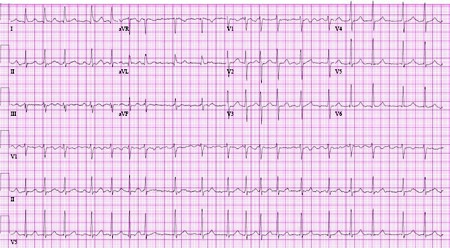
Practical tip
Always suspect atrial flutter if there is a narrow complex tachycardia at 150 bpm (or in the range 130-170 bpm).
Examine all the ECG leads carefully for atrial flutter.
Ventricular rate is a fraction of the atrial rate, for example:
2:1 block = 150 bpm
3:1 block = 100 bpm
4:1 block = 75 bpm.
More info: Types of atrial flutter (with ECG examples)
Further classic ECG findings depend on the type of flutter.
Typical form (anticlockwise isthmus-dependent): negatively directed saw-tooth atrial deflections (f waves) in leads II, III, and aVF, and positive deflections in V1 with atrial rates of 250 to 330 bpm.[5][Figure caption and citation for the preceding image starts]: Typical atrial flutter with variable (3 to 4:1) blockFrom the collection of Dr K.C. Wu [Citation ends].
[Figure caption and citation for the preceding image starts]: Close-up images of leads V1, II, III, aVF demonstrating the features of typical atrial flutter: positive saw-tooth deflections in lead V1 and negative deflections in leads II, III, aVF (arrows)From the collection of Dr K.C. Wu [Citation ends].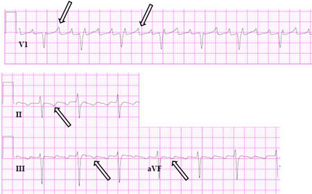
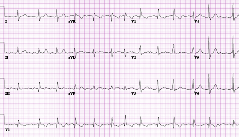
Reverse typical form (clockwise isthmus-dependent): positive flutter waves in leads II, III, and aVF, and negative flutter waves in lead V1.[Figure caption and citation for the preceding image starts]: Reverse typical atrial flutterFrom: Waldo AL. Heart. 2000 Aug;84(2):227-32; used with permission [Citation ends].
 [Figure caption and citation for the preceding image starts]: Selected leads from a patient with reverse typical atrial flutter confirmed at electrophysiological study. The atrial deflections are negative in lead V1 and positive in leads II, III, aVF (arrows)Adapted from: Waldo AL. Heart. 2000 Aug;84(2):227-32; used with permission [Citation ends].
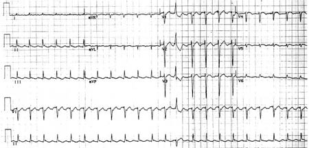
Atypical form (non-isthmus-dependent): continuous undulation of the atrial complex, not meeting criteria for typical or reverse typical flutter, with atrial rates >240 bpm.[2][4][Figure caption and citation for the preceding image starts]: Atypical flutter with right bundle branch blockFrom the collection of Dr K.C. Wu [Citation ends].
 [Figure caption and citation for the preceding image starts]: Close up of leads II, III, V1 showing the continuously undulating pattern of atrial deflections not fitting the criteria for typical or reverse typical atrial flutterFrom the collection of Dr K.C. Wu [Citation ends].
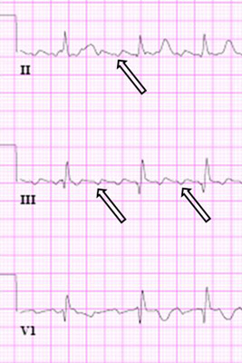
If a patient has atrial flutter with 2:1 block, the diagnosis may not be clear on ECG. Consider use of vagal manoeuvres (such as carotid sinus massage) or intravenous adenosine to confirm the diagnosis, while monitoring the ECG.[5]
If a vagal manoeuvre is performed, this can unmask atrial activity (by causing slowing of AV node conduction and induction of intermittent AV block) and reveal dissociated P waves.[5]
Only use adenosine if deemed necessary for diagnosis and if resuscitation equipment is available.[5]
Consider a 24-hour ambulatory ECG recording. However, tachycardia episodes are usually sporadic and may not be frequent enough to be recorded on ambulatory monitoring.[5]
Blood tests
Always order:
Full blood count[5]
Biochemistry panel, which should include:[5]
Renal function and electrolytes: electrolyte abnormalities are generally not the sole cause of atrial flutter, but imbalances should be checked and corrected
Thyroid function tests: used to rule out thyroid disease as an underlying cause
High-sensitivity troponin: if acute myocardial infarction is suspected. See our topics ST-elevation myocardial infarction and Non-ST elevation myocardial infarction
Digitalis levels: if a patient is taking digitalis drugs (e.g., digoxin).
Consider ordering a D-dimer (± computed tomographic pulmonary angiography) if you suspect pulmonary embolism.
Chest x-ray
Order a chest x-ray; atrial flutter is often associated with a known history of, or suggestive presentation for, lung disease (e.g., pulmonary embolism).
Transthoracic echocardiography (TTE)
Always request a TTE as a baseline investigation to check:[5]
Atrial sizes, ventricular function, and for valvular and pericardial disease
Right ventricular systolic pressures, which may indicate the presence or absence of pulmonary hypertension seen in underlying lung disease.
Further investigations
Pulmonary function tests
Pulmonary function tests are recommended by the European Society of Cardiology if you suspect lung disease as a cause of atrial flutter.[5] However, in practice, other investigations such as high-resolution computed tomography (CT) or CT pulmonary angiography may be used instead.
Electrophysiological studies
Electrophysiology studies require specialist input from an electrophysiologist and are usually indicated to establish the diagnosis of atrial flutter, particularly when catheter ablation is planned.[5]
Electrophysiological studies may also be used if:
Atrial flutter is recurrent, particularly if anti-arrhythmic drug therapy has been ineffective or not tolerated because of adverse effects
The ventricular rate is difficult to control with drugs
Atrial flutter persists despite resolution of the acute underlying illness.
Atrial electrogram
Although the diagnosis of atrial flutter is usually clear on the 12-lead ECG, an atrial electrogram recording (which should be performed by a specialist) can help visualise flutter waves if the diagnosis is uncertain. However, it is rarely used in practice.
Exercise tolerance testing
May be useful if the patient has apparent pre-excitation or a catecholamine-dependent arrhythmia.[5] Can also be considered if there is a history that suggests an exercise-induced arrhythmia.
Myocardial ischaemia testing
Organise myocardial ischaemia testing if the patient has angina, or if you suspect underlying ischaemia (e.g., if significant risk factors for coronary artery disease are present).[5]
Use of this content is subject to our disclaimer