Images and videos
Images


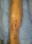
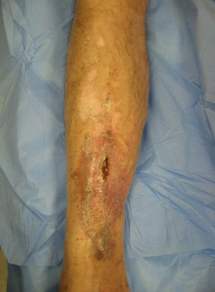
Osteomyelitis
A 62-year-old man suffered an open tibial fracture, which became infected after internal fixation. He continued with intermittent discharge of pus from the front of his tibia for 21 years. Imaging confirmed the presence of chronic osteomyelitis with a central area of dead bone (sequestrum)
Courtesy of the Oxford Bone Infection Unit; used with permission
See this image in context in the following section/s:
Osteomyelitis
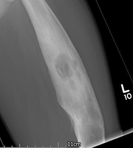
Plain x-ray of the left femur showing a lytic lesion in the medullary canal along with a 'fallen leaf' sign with intramedullary sequestrum noted in the cavity
Courtesy of the Oxford Bone Infection Unit
See this image in context in the following section/s:
Osteomyelitis
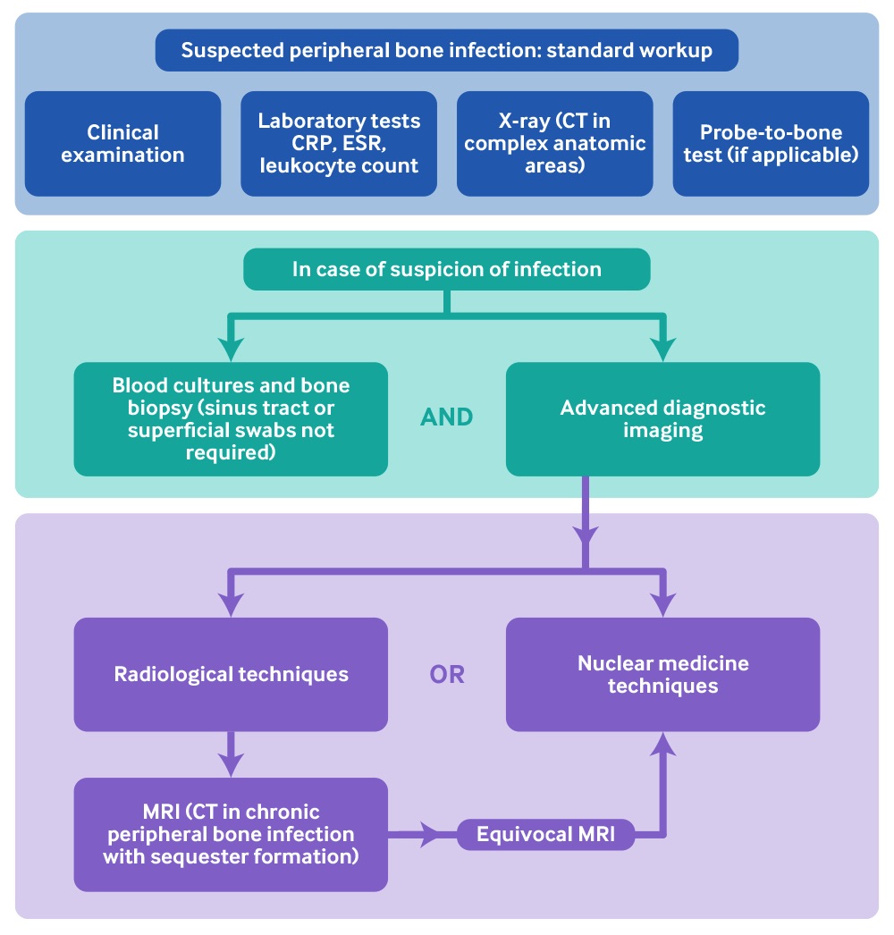
Diagnostic flow chart of peripheral bone infection
Adapted from: Glaudemans AWJM, Jutte PC, Cataldo MA, et al. Consensus document for the diagnosis of peripheral bone infection in adults: a joint paper by the EANM, EBJIS, and ESR (with ESCMID endorsement). Eur J Nucl Med Mol Imaging. 2019;46(4):957-70. http://creativecommons.org/licenses/by/4.0/
See this image in context in the following section/s:
Use of this content is subject to our disclaimer