History and exam
Key diagnostic factors
common
risk factors
In particular, previous osteomyelitis, penetrating injury, intravenous drug misuse, diabetes, HIV infection, recent surgery, distant or local infection, sickle cell disease, rheumatoid arthritis, chronic kidney disease, immunocompromising conditions or, in children, upper respiratory tract or varicella infection.
In children, it is important to know whether they have received the Haemophilus influenzae type B (Hib) vaccination.
limp or reluctance to weight-bear
Common presentation of acute osteomyelitis in children.
non-specific pain at site of infection
Adults with primary or recurrent haematogenous osteomyelitis usually present with vague symptoms consisting of non-specific pain and low-grade fever of 1 to 3 months' duration.[29]
Lumbar vertebral osteomyelitis will present with low back pain and may be associated with intravenous drug use, lower extremity and hip infections, and tuberculosis.
Areas of point tenderness may signify acute or acute on chronic osteomyelitis.
malaise and fatigue
local back pain associated with systemic symptoms
May indicate native vertebral osteomyelitis. Paravertebral muscle tenderness and spasm, and limitation of spine movement, are the predominant physical examination findings.[7]
Pain:
Is localized to the infected disc space area
Is worsened by physical activity or percussion to the affected area
May radiate to the abdomen, hip, leg, scrotum, groin, or perineum
In the lower back may indicate axial osteomyelitis, or lumbar discitis
Spinal cord or nerve root compression and meningitis may occur.
Neurological signs occur more commonly with cervical or thoracic involvement.
paravertebral muscle tenderness and spasm
Associated with limitation of movement in native vertebral osteomyelitis.[7]
local inflammation, tenderness, erythema, or swelling
Differentiate between these signs around the bone, and these signs when associated with a contiguous joint.
In a patient with diabetes, a hot, inflamed foot may be either infection or a Charcot joint. Osteomyelitis may be indicated by:[26]
A local infection with erythema more than 2 cm around an ulcer
A deep foot wound, involving structures deeper than skin and subcutaneous tissues
A chronic foot wound with local infection.
fever
Adults with primary or recurrent haematogenous osteomyelitis usually present with vague symptoms consisting of non-specific pain and low-grade fever of 1 to 3 months' duration.[29]
Fever is only present in up to 45% of patients with bacterial native vertebral osteomyelitis.[7]
Children with acute long bone infection will often have a fever >38°C (>100.4°F).[32]
In the patient with diabetes, a local infection with a temperature of >38°C (>100.4°F) or <36°C (<96.8°F), increased heart rate, or increased respiratory rate may indicate osteomyelitis.[26]
uncommon
spinal cord or nerve root compression
Neurological signs occur more commonly with cervical or thoracic native vertebral osteomyelitis.
Other diagnostic factors
common
wound drainage, acute or old healed sinuses
Persistent drainage and/or sinus tracts are often found adjacent to the area of chronic osteomyelitis and are pathognomonic of infection.[29]
[Figure caption and citation for the preceding image starts]: A 62-year-old man suffered an open tibial fracture, which became infected after internal fixation. He continued with intermittent discharge of pus from the front of his tibia for 21 years. Imaging confirmed the presence of chronic osteomyelitis with a central area of dead bone (sequestrum)Courtesy of the Oxford Bone Infection Unit; used with permission [Citation ends].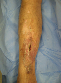
scars, previous flaps, fracture fixation
Evidence of previous surgery suggests chronic osteomyelitis and more complex injury in the original setting.
reduced range of movement
Movement may be restricted due to pain at the site of infection or an associated septic arthritis, particularly in children.
reduced sensation in diabetic foot infection
A patient with diabetes may not report pain due to neuropathy.[26]
Practical tip
The patient with diabetes may not report pain due to neuropathy; they may present only with hyperglycaemia that is difficult to control.
If you can palpate the bone with a probe, this is indicative of osteomyelitis; however, there is no evidence to support the utility of this in diagnosing peripheral bone infection in a patient without diabetes.[2] Be careful with a probe-to-bone test as it can cause harm.
uncommon
urinary tract symptoms
In older adults, the urinary tract may be a source of infection from gram-negative organisms in cases of vertebral osteomyelitis.[37]
torticollis
skin or other infections, recent episode of Staphylococcus aureus bloodstream infection, indwelling catheter
May provide the source of haematogenous or contiguous infection.
A recent history of S aureus bacteraemia associated with new or worsening back or neck pain may indicate native vertebral osteomyelitis.[7]
limb deformity
Angular deformity, or limb shortening, is associated with premature fusion of the physeal plate following childhood osteomyelitis.
tenderness to percussion
May be detected over the subcutaneous border of affected bones in chronic osteomyelitis.
meningitis
May occur in association with native vertebral osteomyelitis.
Use of this content is subject to our disclaimer