Images and videos
Images

Deep vein thrombosis
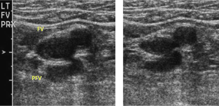
Short-axis ultrasound view showing the femoral vein (FV) and profunda femoris vein (PFV) adjacent to the femoral artery before compression (left) and compressed (right)
From the collection of Jeffrey W. Olin; used with permission
See this image in context in the following section/s:
Deep vein thrombosis
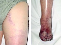
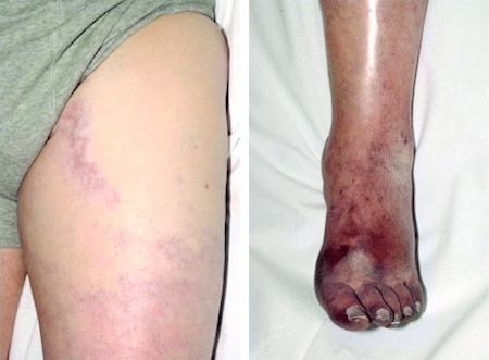
Phlegmasia cerulea dolens: swelling of the left leg and bluish discoloration of the foot
Cooper RM, et al. Phlegmasia cerulea dolens, a rare complication of deep vein thrombosis. Emerg Med J. 2008 Jun;25(6):334
See this image in context in the following section/s:
Use of this content is subject to our disclaimer