Aetiology
The cause of PR has remained elusive after decades of research. However, the following infectious agents have been effectively ruled out: human herpes virus (HHV)-8, cytomegalovirus, Epstein-Barr virus, parvovirus B19, picornaviruses, influenza viruses, parainfluenza viruses, Chlamydia, Legionella, and Mycoplasma.[4][12] There is insufficient evidence to definitively support HHV-6 or HHV-7 as causative agents.[9][12][13] The argument against an infectious cause includes the inability to identify an agent, lack of true epidemics, and inconsistent response to various studied antiviral and macrolide treatments.[12][14]
There is no association of PR with a history of atopy.[4][9] It is unclear if auto-immunity plays a role, but it has been reported that patients with PR are more likely to have a positive antinuclear antibody.[4][9] Cases of PR and PR-like eruptions have been reported in association with coronavirus disease 2019 (COVID-19) infection and following COVID-19 vaccination, though the significance is still being determined.[15][16]
Pathophysiology
While there are no absolute data to support a specific infectious source, most physicians agree that the programmed clinical course, case clustering temporally and among close contacts, low incidence of recurrence (2.8% of individuals experience a second course), and associated prodromal symptoms support an unidentified viral cause.[3][4] Inflammatory changes (increased B lymphocytes, decreased T lymphocytes, elevated erythrocyte sedimentation rate) are seen in some patients with PR. Human herpes viruses (HHV)-6 and HHV-7 are the most widely postulated and studied viral causes, but evidence is conflicting.[11][13]
Classification
Clinical variants of PR
There are several variants of PR, including inverse, papular, urticarial, vesicular, purpuric/haemorrhagic, gigantea of Darier, limb-girdle, irritata, unilateralis, and localised.[1][2][3][4][5][6][7] These atypical forms of PR account for up to 20% of all cases.[5]
Inverse PR:
More likely to be seen in children and adolescents.[3]
The lesions are concentrated in the skin folds, including the axillae and groin, as well as the face, with less involvement of the trunk.[2][3][6]
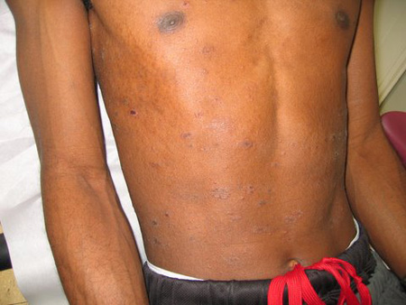
Papular PR:
The herald patch is followed by the eruption of up to hundreds of small maculopapules, some with a scaly border, over the same distribution as classical PR.[1][3][8]
Individuals affected by this variant are more likely to have scalp and facial lesions, and are more likely to report pruritus and post-inflammatory pigmentary alteration.[1][4] The lesions are less likely to be erythematous and more likely to have abundant scale.[1][4][Figure caption and citation for the preceding image starts]: Pityriasis rosea: papular variantFrom the collection of Daniela Kroshinsky, MD, MPH; used with permission [Citation ends].
Urticarial PR:
Characterised by wheal- or hive-like lesions, and is extremely pruritic.[6]
Vesicular PR:
Accounts for less than 1% of all cases of PR.[5]
In this form, vesicles can be limited to the palmoplantar surfaces (resembling dyshidrosis) or generalised (with widespread distribution of lesions).[5] The vesicular lesions are seen most often in combination with the classic papulosquamous lesions.[5]
Most often seen in children and young adults, and can be accompanied by severe itch.[6]
Purpuric/haemorrhagic PR:
This is accompanied by macular purpura without evidence of vasculitis on biopsy.[6]
Palatal petechiae can also be seen in this form.[6]
PR gigantea of Darier:
A very rare form of PR characterised by enormous plaques.[6]
Limb-girdle PR:
In this form, the lesions are concentrated on the shoulders or hips.[6]
PR irritata:
These cases have classic lesions of PR in typical or atypical locations, accompanied by severe pain, burning, or pruritus.[4][7]
PR unilateralis/localised:
PR unilateralis produces an asymmetrical concentration of lesions over one side of the body, while localised PR concentrates the lesions in one body area, in the neck or lower abdomen.[6][7]
Palmoplantar PR:
Demonstrates involvement of the distal extremities, including the palms or soles, with erythematous plaques, and can be vesicular.
Secondary syphilis must be excluded.[9]
Use of this content is subject to our disclaimer