Images and videos
Images


Melanoma
Bluish-white veil of a melanoma
From the personal collection of Dr Hobart Walling and Dr Brian Swick.
See this image in context in the following section/s:
Melanoma
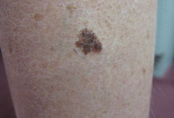
Superficial spreading melanoma
From the personal collection of Dr Hobart Walling and Dr Brian Swick.
See this image in context in the following section/s:
Melanoma
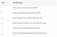
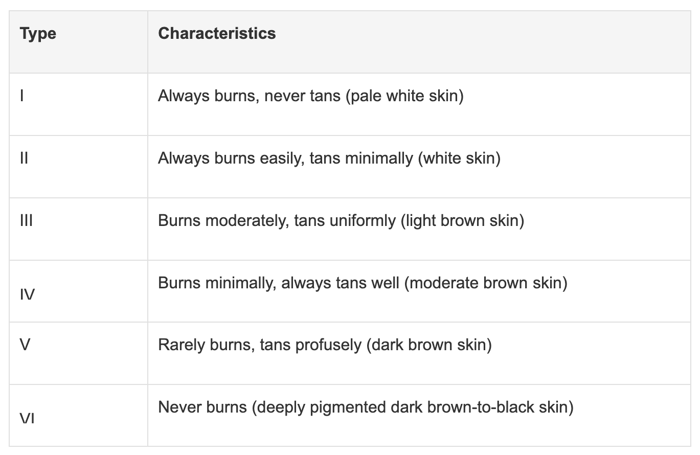
Fitzpatrick skin type
Created by BMJ Knowledge Centre
See this image in context in the following section/s:
Melanoma
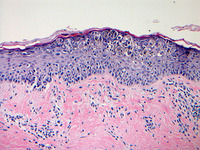
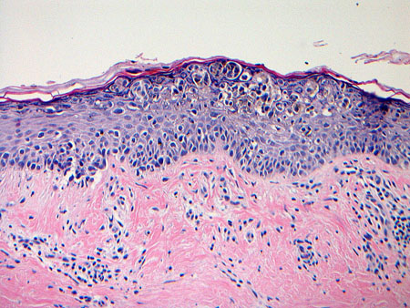
Photomicrograph of melanoma in situ
From the personal collection of Dr Hobart Walling and Dr Brian Swick.
See this image in context in the following section/s:

Melanoma
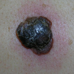
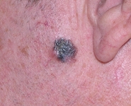
Nodular melanoma
From the personal collection of Dr Hobart Walling and Dr Brian Swick.
See this image in context in the following section/s:

Melanoma
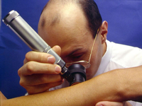
Dermoscopy: the most important application of dermoscopy is distinguishing melanoma from benign melanocytic lesions
© DermNet New Zealand; used with permission
See this image in context in the following section/s:

Melanoma
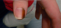
Subungual melanoma in situ
From the personal collection of Dr Hobart Walling and Dr Brian Swick.
See this image in context in the following section/s:

Melanoma
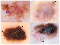
Key dermoscopic features of melanoma: (A) Melanoma presenting with atypical globules and dots of different sizes and shapes (yellow arrows), patches of atypical network (blue arrowhead) and a blue-white veil (blue arrow). (B) Melanoma with diffuse polymorphous vasculature, consisting of serpentine, dotted, and glomerular vessels, can be found throughout the lesion (yellow arrowheads); in addition, patches of atypical network (blue arrowheads) are seen. (C) Superficial spreading melanoma with pseudopods distributed asymmetrically around the lesion (black arrowheads). (D) Melanoma with the regression structure blue-grey peppering (black star); shiny white lines are also seen throughout the entire lesion (red arrows) along with a central blue-white veil (red arrowhead)
Wolner ZJ et al. Enhancing skin cancer diagnosis with dermoscopy. Dermatol Clin. 2017 Oct;35(4):417-37; used with permission
See this image in context in the following section/s:
Melanoma
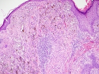
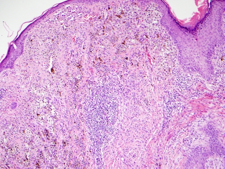
Photomicrograph of Clark's level IV invasive melanoma
From the personal collection of Dr Hobart Walling and Dr Brian Swick.
See this image in context in the following section/s:
Use of this content is subject to our disclaimer