Images and videos
Images


Assessment of dysphagia
Hypertrophic tonsils
From the collection of Dr S. Charous
See this image in context in the following section/s:
Assessment of dysphagia
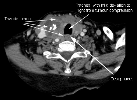
Thyroid tumour compressing cervical oesophagus
From the collection of Dr S. Charous
See this image in context in the following section/s:
Assessment of dysphagia
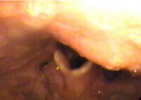
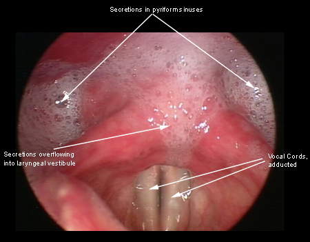
Larynx as a result of cricopharyngeal dysfunction
From the collection of Dr S. Charous
See this image in context in the following section/s:
Assessment of dysphagia
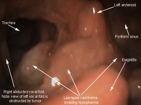
Laryngeal/pharyngeal squamous cell carcinoma
From the collection of Dr S. Charous
See this image in context in the following section/s:

Assessment of dysphagia
Idiopathic achalasia: barium oesophagograms showing a dilated oesophageal body and a tapering stricture in the distal oesophagus ('bird's beak')
From the collection of Dr S. Charous
See this image in context in the following section/s:

Assessment of dysphagia
Endoscopic view of pharynx showing osteophytes pressing inwards on posterior pharyngeal wall, obscuring view of larynx with epiglottis in background
From the collection of Dr S. Charous
See this image in context in the following section/s:

Assessment of dysphagia
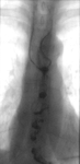
Corkscrew oesophagus from diffuse oesophageal spasm seen on a barium swallow study
From the collection of Dr S. Charous
See this image in context in the following section/s:
Assessment of dysphagia
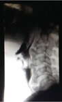
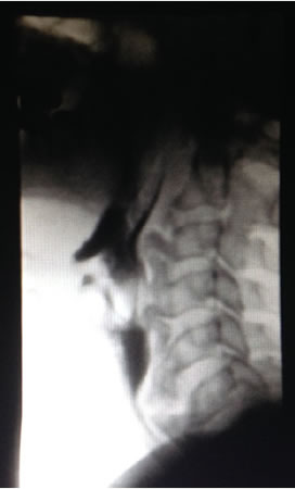
Lateral x-ray during barium swallow, demonstrating osteophytes displacing flow of barium in upper oesophagus
From the collection of Dr S. Charous
See this image in context in the following section/s:

Assessment of dysphagia
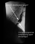
Zenker’s diverticulum: lateral view with barium oesophagram
From the collection of Dr S. Charous, Clinical Professor of Otolaryngology - Head and Neck Surgery, Loyola University Medical Center; used with permission.
See this image in context in the following section/s:
Assessment of dysphagia
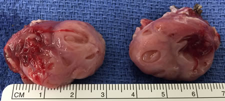
Hypertrophic tonsils
From the collection of Dr S. Charous
See this image in context in the following section/s:
Assessment of dysphagia
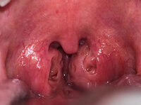
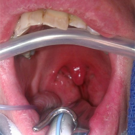
Hypertrophic tonsils causing severely narrowed pharyngeal opening
From the collection of Dr S. Charous
See this image in context in the following section/s:
Use of this content is subject to our disclaimer