Atrial flutter generally results from structural or functional conduction abnormalities of the atria. Structural abnormalities include atrial dilation due to a number of processes (see Risk factors under the Epidemiology section); incisional scars from prior atrial surgery, particularly for congenital heart disease; prior atrial ablation sites; and idiopathic fibrosis within the atrium. In addition, it can be precipitated by toxic and metabolic conditions such as thyrotoxicosis, alcoholism, or pericarditis. Patients taking anti-arrhythmics for chronic suppression of atrial fibrillation may convert to atrial flutter, noted most commonly with Vaughan Williams class Ic drugs (flecainide and propafenone) and amiodarone.[5]Brugada J, Katritsis DG, Arbelo E, et al; ESC Scientific Document Group. 2019 ESC guidelines for the management of patients with supraventricular tachycardia - the Task Force for the management of patients with supraventricular tachycardia of the European Society of Cardiology (ESC). Eur Heart J. 2020 Feb 1;41(5):655-720.
https://academic.oup.com/eurheartj/article/41/5/655/5556821#143629127
http://www.ncbi.nlm.nih.gov/pubmed/31504425?tool=bestpractice.com
[7]Grossman L, Katz M, Beinart R, et al. The clinical outcomes of patients who developed typical atrial flutter on class 1C anti arrhythmic medications treated with hybrid approach. Clin Cardiol. 2019 Jul;42(7):678-83.
https://www.ncbi.nlm.nih.gov/pmc/articles/PMC6605003
http://www.ncbi.nlm.nih.gov/pubmed/31056764?tool=bestpractice.com
[8]Naccarelli GV, Wolbrette DL, Luck JC. Proarrhythmia. Med Clin North Am. 2001 Mar;85(2):503-26;xii.
http://www.ncbi.nlm.nih.gov/pubmed/11233957?tool=bestpractice.com
Occasionally, it can be congenital.[9]Olgin JE, Zipes DG. Libby: Braunwald's heart disease: a textbook of cardiovascular medicine. 8th ed. Philadelphia, PA: Saunders Elsevier; 2007.
The typical form of atrial flutter is a prototypic macro re-entrant arrhythmia, in which the re-entrant wavefront travels up the interatrial septum and down the right atrial free wall ('typical' form) or vice versa ('reverse typical' form). The lateral anatomical boundaries are critical to the development and maintenance of the circuit. One of these boundaries is the tricuspid valve annulus and is fixed or anatomical. The other is generally a functional line of block between the venae cavae.[10]Waldo AL. The interrelationship between atrial fibrillation and atrial flutter. Prog Cardiovasc Dis. 2005 Jul-Aug;48(1):41-56.
http://www.ncbi.nlm.nih.gov/pubmed/16194691?tool=bestpractice.com
Atypical flutters are seen when the cavotricuspid isthmus is not part of the circuit, and are characterised by a continuously undulating pattern by ECG that does not fit strict criteria for the typical and reverse typical forms of atrial flutter. This is a limitation in the mechanistic/anatomical classification scheme, as the exact mechanism can be determined only by electrophysiological mapping studies and not from the ECG alone. Clinical presentations and electrophysiological features of atypical atrial flutter and other types of atrial tachycardia can overlap.[3]Saoudi N, Cosío F, Waldo A, et al. A classification of atrial flutter and regular atrial tachycardia according to electrophysiological mechanisms and anatomical bases; a statement from a joint expert group from the Working Group of Arrhythmias of the European Society of Cardiology and the North American Society of Pacing and Electrophysiology. Eur Heart J. 2001 Jul;22(14):1162-82.
https://academic.oup.com/eurheartj/article/22/14/1162/494426
http://www.ncbi.nlm.nih.gov/pubmed/11440490?tool=bestpractice.com
[Figure caption and citation for the preceding image starts]: Left panel: atrial activation in typical atrial flutter (AFL). Right panel: activation in reverse typical AFL. The atria are represented schematically in a left anterior oblique view, from the tricuspid (left) and mitral rings. The endocardium is shaded and the openings of the superior (SVC) and inferior vena cava (IVC), coronary sinus (CS), and pulmonary veins (PV) are shown. The direction of activation is shown by arrows. Dashed areas mark approximate location of zones of slow conduction and block. Lettering on the right-hand panel marks the low (LPS), mid (MPS), and high (HPS) posteroseptal wall, respectivelyFrom: Waldo AL. Heart. 2000;84:227-227; used with permission [Citation ends].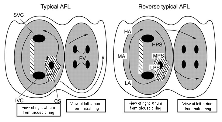
Atrial flutter is traditionally defined according to the ECG appearance, which shows continuous regular electrical activity, most commonly a saw-tooth pattern.[5]Brugada J, Katritsis DG, Arbelo E, et al; ESC Scientific Document Group. 2019 ESC guidelines for the management of patients with supraventricular tachycardia - the Task Force for the management of patients with supraventricular tachycardia of the European Society of Cardiology (ESC). Eur Heart J. 2020 Feb 1;41(5):655-720.
https://academic.oup.com/eurheartj/article/41/5/655/5556821#143629127
http://www.ncbi.nlm.nih.gov/pubmed/31504425?tool=bestpractice.com
It is a macro re-entrant atrial tachycardia with constant P wave/flutter morphology with an atrial rate usually >250 beats per minute (bpm).[5]Brugada J, Katritsis DG, Arbelo E, et al; ESC Scientific Document Group. 2019 ESC guidelines for the management of patients with supraventricular tachycardia - the Task Force for the management of patients with supraventricular tachycardia of the European Society of Cardiology (ESC). Eur Heart J. 2020 Feb 1;41(5):655-720.
https://academic.oup.com/eurheartj/article/41/5/655/5556821#143629127
http://www.ncbi.nlm.nih.gov/pubmed/31504425?tool=bestpractice.com
It is distinguishable from focal atrial tachycardia, which has discrete P waves with an intervening isoelectric segment. Focal atrial tachycardia is caused mechanistically by micro re-entry or increased automaticity and generally has atrial rates in the range of 100 to 250 bpm.[2]Page RL, Joglar JA, Caldwell MA, et al. 2015 ACC/AHA/HRS guideline for the management of adult patients with supraventricular tachycardia. J Am Coll Cardiol. 2016 Apr 5;67(13):e27-115.
https://content.onlinejacc.org/article.aspx?articleid=2443667
http://www.ncbi.nlm.nih.gov/pubmed/26409259?tool=bestpractice.com
Cavotricuspid isthmus-dependent (typical atrial flutter):[5]Brugada J, Katritsis DG, Arbelo E, et al; ESC Scientific Document Group. 2019 ESC guidelines for the management of patients with supraventricular tachycardia - the Task Force for the management of patients with supraventricular tachycardia of the European Society of Cardiology (ESC). Eur Heart J. 2020 Feb 1;41(5):655-720.
https://academic.oup.com/eurheartj/article/41/5/655/5556821#143629127
http://www.ncbi.nlm.nih.gov/pubmed/31504425?tool=bestpractice.com
Anticlockwise atrial flutter with ECG flutter waves characterised by:[2]Page RL, Joglar JA, Caldwell MA, et al. 2015 ACC/AHA/HRS guideline for the management of adult patients with supraventricular tachycardia. J Am Coll Cardiol. 2016 Apr 5;67(13):e27-115.
https://content.onlinejacc.org/article.aspx?articleid=2443667
http://www.ncbi.nlm.nih.gov/pubmed/26409259?tool=bestpractice.com
[5]Brugada J, Katritsis DG, Arbelo E, et al; ESC Scientific Document Group. 2019 ESC guidelines for the management of patients with supraventricular tachycardia - the Task Force for the management of patients with supraventricular tachycardia of the European Society of Cardiology (ESC). Eur Heart J. 2020 Feb 1;41(5):655-720.
https://academic.oup.com/eurheartj/article/41/5/655/5556821#143629127
http://www.ncbi.nlm.nih.gov/pubmed/31504425?tool=bestpractice.com
Negative deflection in leads II, III, aVF
Positive deflection in lead V1
Clockwise atrial flutter (reverse typical atrial flutter) with ECG flutter waves characterised by:
Positive deflection in leads II, III, aVF
Negative deflection in lead V1.
Non-cavotricuspid isthmus-dependent (atypical atrial flutter):[2]Page RL, Joglar JA, Caldwell MA, et al. 2015 ACC/AHA/HRS guideline for the management of adult patients with supraventricular tachycardia. J Am Coll Cardiol. 2016 Apr 5;67(13):e27-115.
https://content.onlinejacc.org/article.aspx?articleid=2443667
http://www.ncbi.nlm.nih.gov/pubmed/26409259?tool=bestpractice.com
Re-entry that does not depend upon conduction through the cavotricuspid isthmus
Circuit is typically defined by atrial scars due to prior heart surgery, ablations, or idiopathic causes
Location determines ablation approach and risks
Multiple sites of re-entry may be present
Can occur in both the left and right atria.