Approach
Many forms of EPTB are paucibacillary, and the diagnosis of EPTB is therefore challenging. Acid-fast bacilli (AFB) smear of biological specimens is often negative. Constitutional symptoms associated with EPTB, (such as fever, weakness, and weight loss) may be infrequent and non-specific. In addition, EPTB is less common than pulmonary TB and may be less familiar to clinicians.
A high level of suspicion is important in evaluating a patient with presence of risk factors (for full details please refer to risk factor section). The firm diagnosis of TB requires culturing of Mycobacterium tuberculosis and this is also important for drug-susceptibility testing. Appropriate specimens are obtained and tested microbiologically and histologically.[41] Although culture remains the diagnostic standard, it can take up to 8-10 weeks using a solid media, and in 10% to 15% of patients the diagnosis of TB is based on clinical grounds. Delays in diagnosis and initiation of therapy are associated with increased mortality.[41]
Rapid diagnostic tests (e.g., nucleic acid amplification tests [NAATs]) are available and can be useful in many settings; some are endorsed by the World Health Organization (WHO) and can detect resistance to some TB drugs.[42]
Tests for all suspected EPTB
As the lungs may be involved in patients with EPTB, sputum for AFB smear and culture is indicated for all suspected patients.[43] Culture-positive sputum becomes useful when the specimens from extrapulmonary sites are culture-negative, and it may also add further information on the infectiousness of the patient.[41] Chest x-ray should be part of the basic initial work-up and may show evidence of active or old TB. A positive TST or IGRA are helpful for diagnosis, but a negative TST or IGRA do not rule out active TB disease. A full blood count should be sent and may show abnormalities.[41]
If the suspicion of TB is high or the patient is very ill, consideration can be given to starting antituberculous medicines as soon as diagnostic specimens are obtained.
Several rapid NAATs, for example, polymerase chain reaction (PCR) assays, are available for the diagnosis of TB, and some are also able to detect resistance to some TB drugs. Although NAATs were originally designed and approved for respiratory specimens, they may also be requested on specimens from other sites where involvement of TB is suspected (e.g., cerebrospinal fluid, lymph node aspirate, lymph node biopsy, pleural fluid, peritoneal fluid, pericardial fluid, synovial fluid or urine).[42] In the US, use of NAATs for extrapulmonary specimens is not approved by the US Food and Drug Administration, and use would be off-label.
Xpert MTB/RIF and Xpert Ultra are rapid NAATs recommended by WHO as initial diagnostic tests in adults and children with signs and symptoms of EPTB.[38][42]
They are also recommended by WHO for detection of rifampicin resistance.[42] Cochrane reviews of Xpert MTB/RIF and Xpert Ultra found that sensitivity of the tests in diagnosing EPTB in patients with presumed infection varied across different site specimens, but the specificity was high.[44][45]
[ ![]() ]
Line probe assays (LPAs) are strip-based tests that can detect TB and determine drug resistance profiles. LPAs are recommended by WHO only for detecting resistance to anti-TB drugs.[42]
]
Line probe assays (LPAs) are strip-based tests that can detect TB and determine drug resistance profiles. LPAs are recommended by WHO only for detecting resistance to anti-TB drugs.[42]
Lateral flow tests that detect lipoarabinomannan (LAM) antigen in urine have emerged as potential point-of-care tests. One Cochrane review found the lateral flow urine lipoarabinomannan (LF-LAM) assay to have a sensitivity of 42% in diagnosing TB in HIV-positive individuals with TB symptoms, and 35% in HIV-positive individuals not assessed for TB symptoms.[46] The WHO recommends that LF-LAM can be used to assist in the diagnosis of active TB in HIV-positive adults, adolescents, and children.[42] This approach is supported by another Cochrane review, which found reductions in mortality and an increase in treatment initiation with use of LF-LAM in inpatient and outpatient settings.[47] Culture would still be required for drug susceptibility testing (DST).
It is recommended that all patients with TB have an HIV test within 2 months of diagnosis. Around 6% of patients with TB are living with HIV.[5]
HIV infection and its treatment may alter the treatment of TB; treatment of HIV may be crucial to the morbidity of HIV-infected TB patients.[48]
TB lymphadenitis
Patients most commonly present with enlarged lymph nodes in the cervical or supraclavicular areas that may be unilateral or bilateral. Scrofula is a term applied to TB adenitis in the neck.
If a patient with superficial lymphadenitis is suspected of having TB, the first diagnostic test is fine-needle aspiration, especially if the lymph node is fluctuant. In addition to AFB smear and culture, aspirate should be submitted for NAAT.[44] If the diagnosis remains in question, a surgical consultation is obtained for lymph node excision.
If the patient has mediastinal lymphadenitis, biopsy is obtained with endobronchial ultrasound (EBUS) bronchoscopy, mediastinoscopy, or thoracoscopy.
Pleural TB
Pleural TB usually presents with symptoms such as pleuritic chest pain, cough, and fever, and a chest x-ray showing a unilateral effusion. The effusion is commonly small to moderate in size; bilateral TB effusions are rare and associated with disseminated disease.
In addition to a chest x-ray, and sputum mycobacterial cultures, a thoracentesis should be performed. Chest x-ray may show no obvious parenchymal disease in 50% of patients with pleural TB. Among those without definite parenchymal involvement, sputum AFB smears are almost always negative and cultures are positive in 20% to 30% of patients. False-negative TST/IGRA are also common.[2]
Pleural fluid analysis is performed on the sample obtained from thoracentesis. Pleural fluid is sent for AFB smear and culture, cell count with differential, protein, LDH, glucose, and pH. AFB smear is rarely positive. Pleural fluid analysis usually shows an exudative effusion that is lymphocyte-predominant and often has low glucose level. The adenosine deaminase (ADA) level may be measured because it is often elevated in pleural TB (sensitivity and specificity approximately 90%), although pneumonia and malignancy, which are frequently differential diagnoses, may also elevate the ADA level.[49] When the ADA level is very low, pleural TB is unlikely. Measurement of pleural fluid free interferon gamma levels is also recommended in diagnosing pleural TB.[41]
Although results of pleural fluid analysis may be helpful, they will seldom confirm a diagnosis of pleural TB. Because a malignancy may also cause a lymphocyte-predominant exudative effusion, the diagnosis of pleural TB is based on microbiology, pathology, identification of granulomas, and negative cytology for malignancy. It is important to obtain a TB isolate for susceptibility testing. Therefore, closed pleural biopsy is indicated when the patient has a lymphocyte-dominant exudative effusion, or even at the same time as thoracentesis if clinical suspicion for TB is very high. The combination of AFB culture and histology from pleural biopsy is the most sensitive to diagnose pleural TB. If results of biopsy are non-diagnostic, thoracoscopy or thoracotomy may be indicated.[50][51]
Skeletal TB (bone and joint TB)
Pain of the involved area is the most common complaint in skeletal TB; constitutional symptoms are usually absent. Diagnosis is based on tissue biopsy. Onset of pain is gradual (over weeks to months) and diagnosis is frequently delayed. Local swelling and limitation of movement may be present. Cold abscesses (non-tender) with sinus tracts may form.
If skeletal TB is suspected, magnetic resonance imaging (MRI) (especially in spinal involvement) or computerised tomography (CT) is obtained. One half of cases will have abnormalities on chest x-ray consistent with TB.[52] Microbiological confirmation of TB is also essential. AFB smears are unlikely to be positive due to low bacillary loads. Cold abscesses, if present, may be aspirated for AFB smear and culture. CT-guided biopsy in vertebral TB will have positive microbiological or histological yields in 65% to 90% of patients.
Synovial biopsy should be done to diagnose TB arthritis. Biopsy may yield culture positive in 90% to 95%, and should be performed if the diagnosis of TB arthritis remains in question.[16] In joint involvement, evaluation of synovial fluid is usually not diagnostic; WBC counts in TB arthritis are usually 10,000-20,000/mL but can be much higher. AFB smear is positive in <20% but culture may be positive in up to 80%.
Central nervous system TB
Central nervous system (CNS) TB may present with meningitis or intracranial tuberculomas. Diagnosis of TB meningitis is dependent upon CSF examination, and its rapid diagnosis is essential for improved outcomes. When there is high pre-test probability for CNS TB, empirical TB treatment should be started while awaiting microbiological confirmation.
Signs and symptoms of meningeal TB include headache, neck stiffness, altered mental status, and cranial nerve abnormalities. Only 38% of children with TB meningitis have fever and 9% report photophobia. Seizures are common in children and older people.
In the presence of meningeal signs, the patient undergoes lumbar puncture and the CSF is submitted for cell count with differential, glucose, protein, AFB smear and culture, Gram stain, and bacterial culture. PCR can be added if available. The usual results of analysis include a lymphocyte predominance, elevated protein, and reduced glucose. ADA levels may be useful in the diagnosis of CNS TB.[41] Although smears of spinal fluid are frequently negative, the diagnostic yield is dependent on the volume of CSF submitted and the quality of examination.[53]
In order to maximise the sensitivity of TB diagnosis by spinal fluid analysis, some experts suggest increased CSF volume (≥6 mL of spinal fluid for AFB) and repeated sampling (up to three lumbar punctures on different days).[53][54]
How to perform a diagnostic lumbar puncture in adults. Includes a discussion of patient positioning, choice of needle, and measurement of opening and closing pressure.
AFB culture is the definitive standard for diagnosis but treatment must not wait until culture results are available. Treatment is initiated presumptively based on clinical suspicion, risk factors, and CSF results.[16]
Head CT or MRI may show oedema, hydrocephalus, basilar meningeal thickening, or tuberculomas. Tuberculomas present as a slowly growing focal lesion, or, rarely, with signs and symptoms consistent with increased intracranial pressure. CSF analysis is usually normal and diagnosis is based on CT or MRI findings.
Up to 50% of patients have chest x-ray abnormalities consistent with pulmonary TB.[24]
Abdominal TB
Abdominal TB includes TB peritonitis and TB of the gastrointestinal tract. Chest x-ray may show evidence of old or concurrent pulmonary TB. Definitive diagnosis is based on culture growth of M tuberculosis from ascitic fluid or a biopsy of the lesion. Patients may have disease for months before the diagnosis is made. Peritoneal disease is the more common presentation. The presenting symptoms include abdominal swelling, abdominal pain, fever, and change in bowel habits. In TB enteritis (TB of the gastrointestinal tract), common sites of involvement are the ileocecal and anorectal areas. Chronic abdominal pain is the most common symptom in addition to changes in bowel habits and haem-positive stool. Patients may develop small bowel obstruction or a right lower quadrant mass.
CT scan of the abdomen, ascitic fluid analysis, and peritoneal biopsy are done initially.
CT scan may show ascites, bowel-wall thickening, or abdominal lymphadenopathy.
Ascitic fluid analysis is non-specific and rarely AFB smear-positive. Although the sensitivity of culture from peritoneal fluid is high (92%), results require up to 8 weeks and delay in initiating treatment is associated with higher mortality.
Peritoneal biopsy (laparoscopy or laparotomy) is the most effective means for diagnosis. Direct inspection may reveal miliary nodules over the peritoneum and allow a presumptive diagnosis in 80% to 95%. Biopsy demonstrates caseating granulomas (up to 100%) and the presence of AFBs on examination in 67% of samples.[15][61] Ascitic fluid ADA and free interferon gamma levels may have a role in diagnosing abdominal TB.[41]
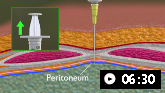
Demonstrates how to perform diagnostic and therapeutic abdominal paracentesis.
Abdominal ultrasound may also aid in diagnostic evaluation but should not be used alone for TB diagnosis.[67] [
Colonoscopy and biopsy are carried out to diagnose TB enteritis. Colonoscopy will reveal ulcers, pseudopolyps, or nodules. Definitive diagnosis is based on biopsy, which usually shows granulomas and culture positive for TB.[15]
Genitourinary TB
Chest x-ray is abnormal in 40% to 75% of patients with genitourinary (GU) TB.
Diagnosis relies on culturing M tuberculosis from morning urine samples (collection of three specimens is recommended) or biopsy of the lesion. The common symptoms are dysuria, haematuria, and urinary frequency. Symptoms may be absent in 20% to 30% of patients. Genital TB in men may present as a scrotal mass, whereas in women it may be asymptomatic or cause pelvic pain, menstrual disorders, or infertility. Constitutional symptoms are rare. Extensive renal destruction may have occurred by the time GU TB is diagnosed.[34][68]
Urinalysis is done initially. Results commonly show pyuria, haematuria, or proteinuria, although they may be normal. While the pyuria is classically described as 'sterile', superimposed bacterial infections may be present in patients with GU TB. Urine culture for TB may be positive in 80% of patients; three samples for culture improve sensitivity. NAATs of the urine can be helpful adjunctive tools for the rapid diagnosis of GU TB.[44] Definitive diagnosis of genital TB is based on tissue biopsy.[68]
Pericardial TB
Chest x-ray shows cardiomegaly (in 70% to 95% of cases) and pleural effusion (in about 50%). ECG is low voltage (in about 25%) and shows T-wave inversion (in about 90%). Echocardiography, CT, or MRI shows pericardial effusion and thickness across the pericardial space. Diagnosis of pericardial TB requires aspiration of pericardial fluid (by pericardiocentesis) or, usually, pericardial biopsy. Pericardial fluid is exudative with increased leukocytes, predominantly lymphocytes. Pericardial fluid should be sent for AFB smear (sensitivity 0% to 42%), culture (sensitivity 50% to 65%), and ADA. The sensitivity and specificity of an elevated ADA level in pericardial fluid (at a threshold of 40 U/L) are 88% and 83%, respectively. A positive AFB smear and elevated ADA level suggests TB pericarditis; positive culture confirms diagnosis of TB pericarditis.[41] Haemorrhagic effusion is often seen. Pericardial biopsy offers a higher diagnostic yield. Pericardial tissue should be sent for histological examination (sensitivity 73% to 100%) and culture.[41]
Disseminated TB
The diagnosis of disseminated TB concentrates on the organs most likely to be involved. The most commonly involved organs (in order) are lungs, liver, spleen, kidneys, and bone marrow. Patients with disseminated TB will typically have constitutional symptoms including fever (90%), anorexia (78%), and sweats (76%).
If disseminated TB is suspected, chest x-ray (if non-diagnostic, consider a chest CT), sputum for AFB smear and culture, blood culture for mycobacteria, and first-morning-void urine for AFB are obtained; lumbar puncture and biopsy of superficial lymph nodes are also done if applicable. Sputum smear will be positive in one-third of patients with culture positive in about 60%.
As delays in treatment are associated with increased mortality, a rapid diagnostic test (i.e., faster than culture results) is frequently needed. If sputum smears are negative and chest x-ray is abnormal, bronchoscopy with transbronchial biopsies are indicated. If results are non-diagnostic, bone marrow or liver biopsy is also done. Both have similar sensitivities, but bone marrow biopsy may be preferred because of its lower procedure risk. If thrombocytopenia or leukopenia are present, the sensitivity of bone marrow biopsy is increased.[16][69][70][71]
Tests for latent TB infection (LTBI)
Investigations for LTBI (also sometimes referred to as TB infection) in a person exposed to M tuberculosis but without signs of active TB are based on the tuberculin skin test (TST) or interferon gamma release assays (IGRAs). The TST and IGRA measure the response of T cells to TB antigens. These tests have limited use in active TB infection and should not be used alone to exclude a diagnosis of active TB.
The TST uses intra-dermal injection of purified protein derivative to evaluate for delayed hypersensitivity response in order to diagnose prior exposure to TB. Different cut-offs in size of induration are used to define a positive test, depending on the patient's risk factors. Response to TST may be diminished in patients with factors such as HIV infection or poor nutrition.[72] IGRAs measure the release of interferon-gamma from T cells reacting to TB antigens.
TB antigen-based skin tests (TBSTs) are a new class of tests that have been developed to measure the cell-mediated immunological response to M tuberculosis specific antigens. The WHO recommends that TBSTs may be used to test for LTBI, reporting that the diagnostic accuracy of TBSTs is similar to that of IGRAs and greater than that of the TST.[73]
Use of this content is subject to our disclaimer