Aetiology
Physiological or pathophysiological stimuli that cause vasopressin release combined with fluid intake may lead to hyponatraemia. Additionally, reduced thyroid function and/or adrenal insufficiency may contribute to an increased effect, or release, of vasopressin.
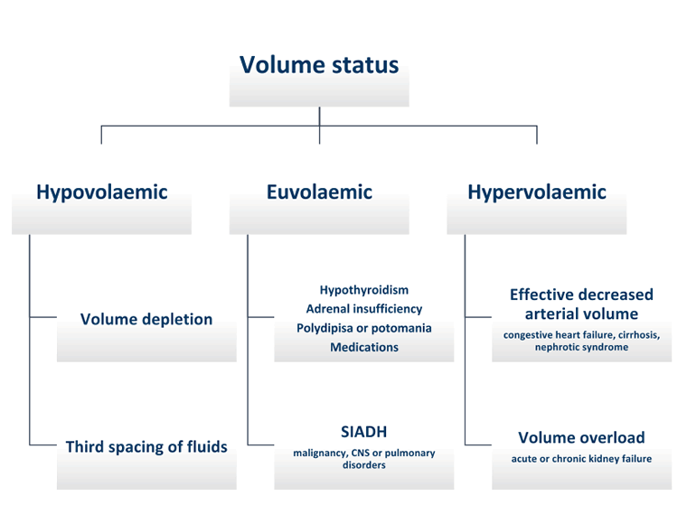
Physiological stimuli for vasopressin release include loss of intravascular volume (hypovolaemic hyponatraemia) and the loss of effective intravascular volume (hypervolaemic hyponatraemia).[2][3][12]
Causes of hypovolaemic hyponatraemia include:[1][2]
Gastrointestinal fluid loss (e.g., severe diarrhoea or vomiting)
Third spacing of fluids (e.g., pancreatitis, severe hypoalbuminaemia)
Salt-wasting nephropathy
Cerebral salt-wasting syndrome (a rare cause of hyponatraemia resulting from urinary salt wasting; elevated brain natriuretic peptide has been implicated)
Mineralocorticoid deficiency
Causes of hypervolemic hyponatraemia include:[1][2]
Acute kidney injury/chronic kidney disease (low sodium levels in advanced kidney disease or dialysis patients is due to relatively higher water versus salt intake with poor excretion due to underlying kidney disease)
Congestive heart failure
Cirrhosis
Nephrotic syndrome
In pregnancy, physiological hypervolaemia leads to a mild dilutional hyponatraemia, which can be exacerbated by excessive fluid intake or IV fluid administration at the time of labour and delivery.[13] Normal sodium levels in pregnancy are 130-140mEq/L; sodium levels lower than 130mEq/L should be investigated for an alternative underlying cause.[14]
Non-osmotic, pathological vasopressin release can also occur in the setting of normal volume status, as is the case with euvolaemic hyponatraemia. The main causes of euvolaemic hyponatraemia include:[1][3]
Medications (e.g., vasopressin, diuretics, antidepressants, opioids). The most common are thiazide diuretics and antidepressants.[15]
Syndrome of inappropriate antidiuretic hormone: can result from malignancy (e.g., small cell lung cancer, gastrointestinal tract cancers); central nervous system disorders (e.g., subarachnoid haemorrhage, meningitis, encephalitis); pulmonary disease (e.g., pneumonia); or other non-specific causes (e.g., medications, pain, nausea, stress, general anaesthesia). It can also be idiopathic.
High fluid intake: can result from intense/prolonged physical activity (e.g., marathon running, military training, wilderness exploration); surgery; primary polydipsia (also referred to as psychogenic polydipsia); or potomania, which is caused by a low intake of solutes and electrolytes with relatively high fluid intake.[16]
Medical testing: although less common, euvolaemic hyponatraemia related to excessive fluids can occur in the setting of medical testing such as cardiac catheterisation or colonoscopy.
Hypovolaemic and euvolaemic hyponatraemia can also be iatrogenic.
Common causes of hyponatraemia include true volume depletion, effective arterial volume depletion (e.g., congestive heart failure, cirrhosis), and medication-induced hyponatraemia due to thiazide diuretics or antidepressants. For conditions associated with hyponatraemia, the more severe the condition, the greater the likelihood of hyponatraemia. The severity of hyponatraemia may also progress as condition severity increases.
Many drugs have been associated with hyponatraemia, but the most common agents include:[3][17]
Vasopressin analogs: desmopressin, oxytocin
Medications that stimulate vasopressin release or potentiate the effects of vasopressin: selective serotonin-reuptake inhibitors and most other antidepressants, morphine and other opioids[18][19]
Medications that impair urinary dilution: thiazide diuretics[12][15][20]
Medications that cause hyponatraemia by an uncertain mechanism of action: carbamazepine or its analogs, vincristine, nicotine, antipsychotics, chlorpropamide, cyclophosphamide, non-steroidal anti-inflammatory drugs
Illicit drugs: methylenedioxy-methamfetamine (MDMA or ecstasy) causes vasopressin release, and has been associated with acute, life-threatening hyponatraemia
Some causes of hyponatraemia are not directly related to vasopressin release and are therefore not regarded as true hypo-osmolar hyponatraemia. For example, hypertonic hyponatraemia can be caused by hyperglycaemia or the intake of hypertonic fluids, which pull water out of cells (e.g., mannitol, sorbitol); pseudohyponatraemia (also known as isotonic hyponatraemia) is an artifact of incorrect measurement of serum sodium concentrations due to high lipid and/or protein in the plasma.[11] The most common cause of high serum protein levels is multiple myeloma.
[Figure caption and citation for the preceding image starts]: Potential aetiologies of hypotonic hyponatraemia based on volume status of patient. CNS, central nervous system; SIADH, syndrome of inappropriate antidiuretic hormoneProduced by the BMJ Knowledge Centre; adapted from algorithm supplied by Dr J. Veis [Citation ends].
Pathophysiology
Sodium homeostasis is maintained by vasopressin, aldosterone, thirst, and the kidneys. Hyponatraemia occurs when total body sodium concentration falls to <135 mmol/L due to excess water in the body caused by water retention and/or water intake relative to sodium.[2][3] Total body sodium may be low, normal, or elevated.
Vasopressin (arginine vasopressin, also known as antidiuretic hormone) is a hormone that is released from the posterior pituitary and acts on the kidneys to increase water reabsorption. Under normal conditions, if excessive fluid intake occurs, vasopressin release is suppressed, urine osmolality decreases, and the excess water is excreted to return the plasma osmolality to normal.[1] However, in hypo-osmolar hyponatraemia, physiological or pathophysiological factors lead to vasopressin release and/or increased effect, urinary dilution is inhibited, and abnormal water reabsorption occurs that is out of balance with solutes. This lowers the serum osmolality. Vasopressin release is dependent on both responsive serum osmolality and circulating volume (monitored by baroreceptors). Normally, when serum osmolality is high and/or circulating volume is low, vasopressin is released so that water is reabsorbed in the kidneys.[1] However, conditions causing significant intravascular volume depletion can lead to vasopressin release, even in the presence of low serum osmolality. If this occurs, hyponatraemia ensues.
According to the Edelman equation, hyponatraemia develops due to a gain of free water, a loss of serum sodium, or a combination of both.[3]
Cerebral oedema is a medical emergency. If hyponatraemia develops acutely (i.e., <48 hours), the brain does not have time to adapt. Osmolality inside the brain cells remains higher than the serum and water enters the brain cells causing cerebral oedema. This leads to symptoms including nausea, vomiting, altered mental status, and eventually seizures and/or brain herniation and death. With more chronic hyponatraemia, brain adaptation occurs with the loss of intracellular osmolytes, returning brain volume to normal after approximately 48 hours. These patients are generally asymptomatic or present with only mild cognitive symptoms (e.g., confusion, balance difficulties).
Classification
Although there is no definitive classification system, hyponatraemia can be classified according to severity/serum tonicity, time of onset, and/or volume status. Volume status is the most important element in determining the aetiology, while the time of onset is the most important factor in determining the rate of correction and risk of cerebral oedema.
Severity
While there is no consensus on exact serum sodium values, hyponatraemia has been classified according to severity:[2]
Mild: serum sodium = 130-135 mmol/L
Moderate: serum sodium = 125-129 mmol/L
Severe: serum sodium <125 mmol/L
Time of onset
Hyponatraemia can be classified according to its time and speed of onset.[3]
Acute hyponatraemia:
Hyponatraemia that is documented to have occurred over <48 hours. Cerebral oedema occurs more frequently when hyponatraemia develops over <48 hours.[2]
Chronic hyponatraemia:
Hyponatraemia that is documented to have occurred over ≥48 hours.
If hyponatraemia cannot be classified by time of onset, treatment should be based on symptoms.
Serum tonicity
The most common type of hyponatraemia is hypotonic hyponatraemia, while hypertonic and isotonic hyponatraemia are less common.
Hypotonic hyponatraemia:[2]
Hypovolemic hyponatraemia
Hypervolemic hyponatraemia
Euvolemic hyponatraemia
Hypertonic hyponatraemia:
Can be due to intake of hypertonic fluids (e.g., mannitol, sorbitol) or hyperglycaemia-induced hyponatraemia. Hyperglycaemia-induced hyponatraemia may also be isotonic or, rarely, hypotonic.
Isotonic hyponatraemia:
Pseudohyponatraemia.
Volume status
Hypovolemic hyponatraemia:
Occurs when total body water and sodium are both decreased, but total water is repleted in excess of sodium. Low intravascular volume activates baroreceptors, which leads to vasopressin release and water reabsorption. With subsequent water intake, hyponatraemia develops. Associated with significantly low intravascular volume either from fluid losses (e.g., diarrhoea, bleeding, urinary loss) or third spacing of fluids (e.g., pancreatitis, severe hypoalbuminaemia).[2]
Hypervolemic hyponatraemia:
Occurs when total body water and sodium both increase, but total body water increases to a greater extent. Associated with baroreceptor perception of low intravascular volume, which leads to inappropriate vasopressin release with water retention despite overall increases in total body water and sodium. Typically seen in congestive heart failure, cirrhosis with ascites, or nephrotic syndrome.[2]
Euvolemic hyponatraemia:
Occurs when total body water increases, but total body sodium remains unchanged. Associated with pathological vasopressin release, but is not associated with either intravascular volume depletion or hypervolaemia. Can be due to certain medications (e.g., selective serotonin-reuptake inhibitors, thiazide diuretics), or syndrome of inappropriate antidiuretic hormone as a result of pulmonary or central nervous system disorders or malignancy.[2]
Use of this content is subject to our disclaimer