Images and videos
Images


Dental abscess
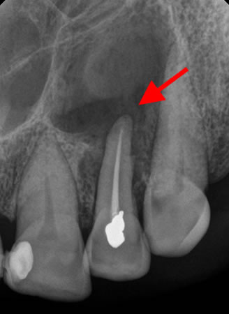
Periapical radiograph showing large periapical radiolucency related to root canal-treated lateral incisor; final pathology was consistent with a periapical cyst
From the personal collection of Melanie S. Lang and Thomas B. Dodson
See this image in context in the following section/s:
Dental abscess
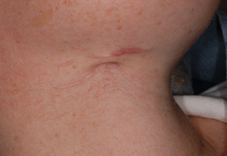
Extra-oral sinus tract in the right anterior neck related to retained necrotic mandibular first molar
From the personal collection of Melanie S. Lang and Thomas B. Dodson
See this image in context in the following section/s:
Dental abscess
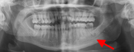
Panoramic radiograph showing generalised advanced horizontal periodontal bone loss with periapical radiolucency (see arrow) related to left mandibular first molar consistent with combined endodontic/periodontal abscess
From the personal collection of Melanie S. Lang and Thomas B. Dodson; used with permission
See this image in context in the following section/s:

Dental abscess
Panoramic radiograph with gutta percha tracking of intra-oral fistula; shows large periapical radiolucency related to failed root canal treatment (note the multiple root canal-treated teeth)
From the personal collection of Melanie S. Lang and Thomas B. Dodson
See this image in context in the following section/s:
Dental abscess
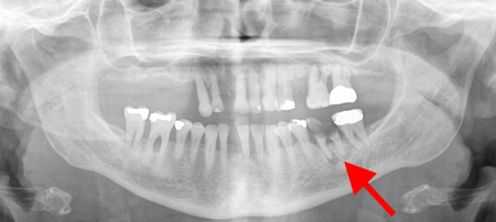
Panoramic radiograph showing periapical abscess related to the lower-left second molar
From the personal collection of Melanie S. Lang and Thomas B. Dodson
See this image in context in the following section/s:

Dental abscess
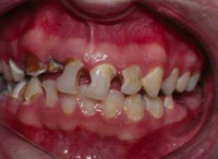
Poor oral hygiene with lack of dental care and resultant rampant decay
From the personal collection of Melanie S. Lang and Thomas B. Dodson
See this image in context in the following section/s:

Dental abscess
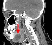
Sagittal CT scan with contrast showing submandibular space abscess
From the personal collection of Melanie S. Lang and Thomas B. Dodson
See this image in context in the following section/s:
Dental abscess
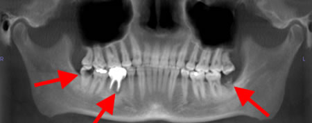
Panoramic radiograph showing decayed bilateral mandibular third molars and failed root canal treatment with periapical lesion related to the right mandibular first molar (middle arrow); also shows carious bilateral mandibular third molars on both sides
From the personal collection of Melanie S. Lang and Thomas B. Dodson
See this image in context in the following section/s:
Use of this content is subject to our disclaimer