Images and videos
Images
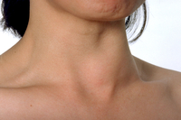
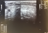

Assessment of thyroid mass
Ultrasound image of thyroid nodule
Courtesy of Getty images; used with permission
See this image in context in the following section/s:
Assessment of thyroid mass
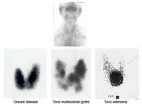
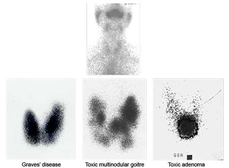
Iodine uptake scans: typical appearances of absent uptake in thyroiditis (top panel); diffuse increased uptake in Grave’s disease (lower left panel); areas of increased and decreased uptake in toxic multinodular goitre (lower middle panel); single area of increased uptake in a toxic adenoma (lower right panel)
Courtesy of Dr Petros Perros
See this image in context in the following section/s:

Assessment of thyroid mass
Nodule sonographic characteristics and threshold size for fine needle aspiration (FNA).
Created by the BMJ Knowledge Centre and Dr B.C. Stack Jr; adapted from Haugen BR, Alexander EK et al. Thyroid 2016 Jan;26(1):1-133
See this image in context in the following section/s:

Assessment of thyroid mass
Goitre
Courtesy of Mediscan/Alamy: used with permission
See this image in context in the following section/s:
Assessment of thyroid mass
TI-RADS calculator: chart showing five categories on the basis of the ACR Thyroid Imaging, Reporting and Data System (TI-RADS) lexicon, TR levels, and criteria for fine-needle aspiration or follow-up ultrasound
Adapted from Tessler FN et al. J Am Coll Radiol 2017 May;14(5):587-95
See this image in context in the following section/s:
Assessment of thyroid mass
Algorithm for the evaluation of a thyroid mass
Created by the BMJ Knowledge Centre and Dr B.C. Stack Jr (FNA, fine needle aspiration; PTH, parathyroid hormone; TSH, thyroid-stimulating hormone)
See this image in context in the following section/s:
Use of this content is subject to our disclaimer