Images and videos
Images


Dermatophyte infections
Tinea pedis. Intense inflammation produces hyperpigmentation and vesicle formation. Vesiculobullous form of tinea pedis
Department of Dermatology Medical University of South Carolina; used with permission
See this image in context in the following section/s:

Dermatophyte infections
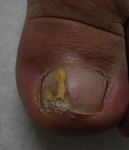
Distal lateral subungual onychomycosis
From the collection of Professor Antonella Tosti; used with permission
See this image in context in the following section/s:
Dermatophyte infections
Infant presenting with rash formerly known as moniliasis, now called candidiasis, caused by Candida spp.
Public Health Image Library, CDC
See this image in context in the following section/s:
Dermatophyte infections
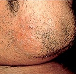
Tinea barbae. Note the pustules in the follicles, redness, and scaling
Department of Dermatology Medical University of South Carolina; used with permission
See this image in context in the following section/s:
Dermatophyte infections
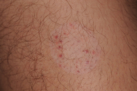
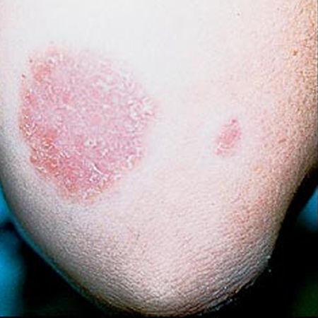
Annular lesion on the elbow, with a silvery scale. No central clearing. Microscopic examination with potassium hydroxide revealed no fungal elements. Despite the resemblance to tinea corporis, there was a similar lesion on the extensor surface of both knees and a family history that together confirmed the diagnosis of psoriasis
Department of Dermatology Medical University of South Carolina; used with permission
See this image in context in the following section/s:

Dermatophyte infections
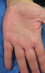
Tinea manuum
From the collection of Professor Antonella Tosti; used with permission
See this image in context in the following section/s:
Dermatophyte infections
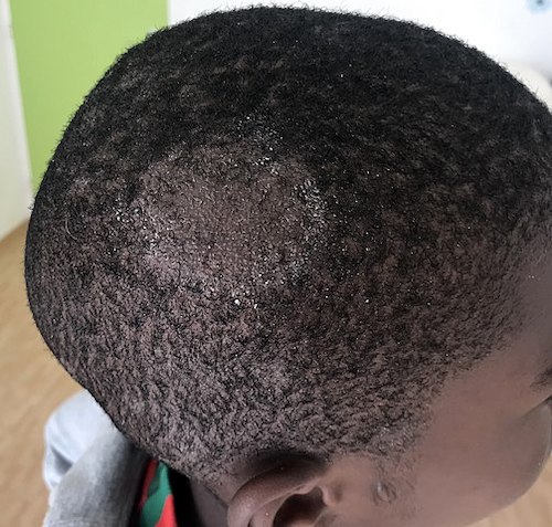
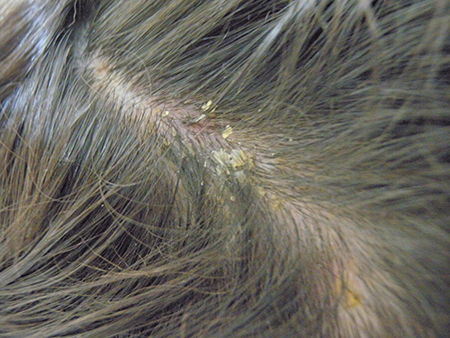
Tinea capitis in a child with Fitzpatrick type VI skin with the typical appearance of fine scale and brown hair, which may be visualised as black dots
Gzzz, Wikimedia Commmons CC-BY-SA-4.0
See this image in context in the following section/s:
Dermatophyte infections
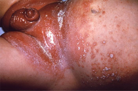
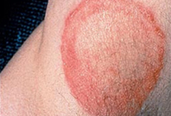
Tinea corporis of the axilla. Central clearing with an active border of inflammation noted. Satellite lesion is present
Department of Dermatology Medical University of South Carolina; used with permission
See this image in context in the following section/s:
Dermatophyte infections
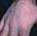
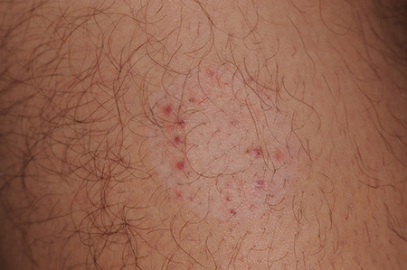
Majocchi granuloma
From the collection of Professor Antonella Tosti; used with permission
See this image in context in the following section/s:

Dermatophyte infections
Tinea manuum. On the extensor surface of the hand there is extensive inflammation, scaling, hyperkeratosis, and erythema
Department of Dermatology Medical University of South Carolina; used with permission
See this image in context in the following section/s:
Dermatophyte infections
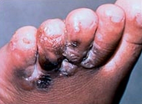
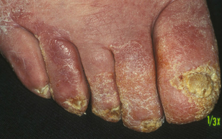
Vesiculobullous form of tinea pedis and onychomycosis
From the collection of Professor Antonella Tosti; used with permission
See this image in context in the following section/s:
Dermatophyte infections
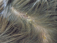
Tinea capitis
From the collection of Professor Antonella Tosti; used with permission
See this image in context in the following section/s:
Dermatophyte infections
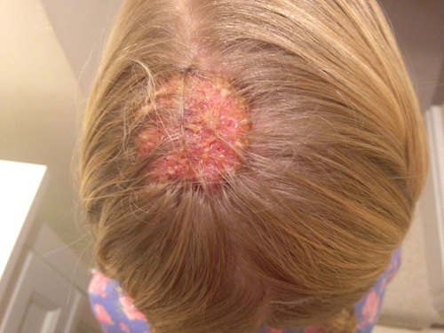
A kerion (abscess due to dermatophyte infection) in a child with Fitzpatrick type 1 skin
Reproduced with permission from Feetham JE, Sargant N. Kerion celsi: a misdiagnosed scalp infection. Arch Dis Child. 2016 May;101(5):503
See this image in context in the following section/s:
Use of this content is subject to our disclaimer