Approach
Dermatophyte infections are typically diagnosed based on clinical assessment; presentation varies according to site of infection.
Laboratory confirmation of diagnosis is helpful in many cases; fungal nail infection must be confirmed by potassium hydroxide (KOH) and culture or periodic acid-Schiff (PAS) staining on a nail clipping prior to treatment.[13]
History
A history of skin, hair, or nail changes, which may or may not cause discomfort, in a characteristic location for days to months is typical.
Patients may have an associated history of:[7][8][9]
Diabetes
Atopy
Exposure to people, farm animals, or pets with skin infections
Exposure to hot, humid weather
Wearing occlusive clothing or footwear
Hyperhidrosis
Use of topical or systemic glucocorticoids or immunosuppressive medications
Immunosuppressive disease, such as HIV.
Onychomycosis may cause local pain, paraesthesias, and difficulties performing activities of daily life. Onychomycosis can predispose to other infections, such as paronychia, tinea pedis, and cellulitis, especially, for example, in people with diabetes.[16]
General examination
General assessment may reveal circumscribed skin lesions of the trunk and inflammatory or scaling lesions of hands, feet, interdigital areas, groin (but sparing the scrotum), and fingernails or toenails.
Skin lesions of the face (tinea faciale), trunk and extremities (tinea corporis), hands (tinea manuum), and groin (tinea cruris) reveal a characteristic pattern of inflammation, termed an active border. The inflammatory response is characterised by a greater degree of redness and scaling at the edge of the lesion, with occasional blister formation. Central clearing of the lesion is often present and distinguishes dermatophytoses from other papulosquamous eruptions such as psoriasis or lichen planus, in which there is a uniform inflammatory response throughout the skin lesion.
[Figure caption and citation for the preceding image starts]: Tinea corporis of the axilla. Central clearing with an active border of inflammation noted. Satellite lesion is presentDepartment of Dermatology Medical University of South Carolina; used with permission [Citation ends].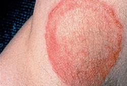 [Figure caption and citation for the preceding image starts]: Annular lesion on the elbow, with a silvery scale. No central clearing. Microscopic examination with potassium hydroxide revealed no fungal elements. Despite the resemblance to tinea corporis, there was a similar lesion on the extensor surface of both knees and a family history that together confirmed the diagnosis of psoriasisDepartment of Dermatology Medical University of South Carolina; used with permission [Citation ends].
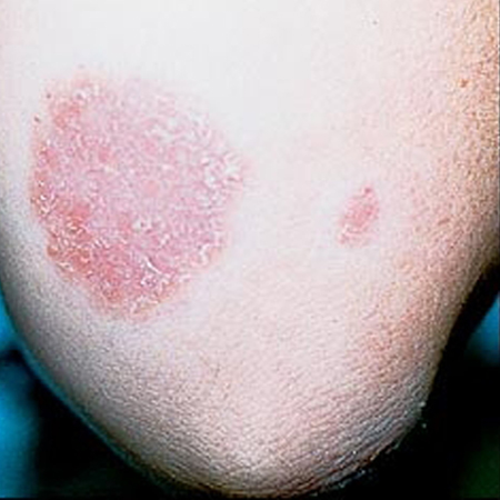
Examination of the scalp
Assessment may reveal hair loss from the scalp or circumscribed areas of scaling and inflammation in the beard area or scalp. Also include palpation of the occipital area and neck for enlarged lymph nodes when examining the scalp.
Non-inflammatory tinea capitis typically presents as fine scaling with single or multiple scaly patches of circular alopecia. Typical dermoscopic findings characteristic of tinea capitis include: black dots, comma hairs (short hairs that bend and grow back toward the scalp, resembling a comma), and corkscrew hairs (short hairs that are coiled up like a corkscrew).[17]
Kerion is a variant of tinea capitis; it is the development of an abscess caused by fungal infection that may also have associated bacterial infection.[18] Swabs should be sent for culture.
[Figure caption and citation for the preceding image starts]: Tinea capitisFrom the collection of Professor Antonella Tosti; used with permission [Citation ends].
[Figure caption and citation for the preceding image starts]: Tinea capitis in a child with Fitzpatrick type VI skin with the typical appearance of fine scale and brown hair, which may be visualised as black dotsGzzz, Wikimedia Commmons CC-BY-SA-4.0 [Citation ends].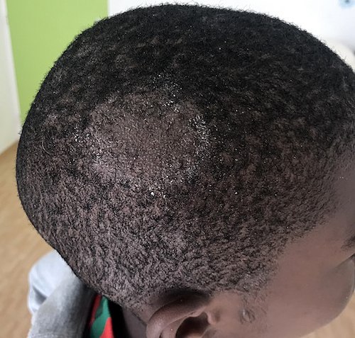
[Figure caption and citation for the preceding image starts]: A kerion (abscess due to dermatophyte infection) in a child with Fitzpatrick type 1 skinReproduced with permission from Feetham JE, Sargant N. Kerion celsi: a misdiagnosed scalp infection. Arch Dis Child. 2016 May;101(5):503 [Citation ends].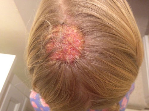
Examination of the face
Tinea barbae involves the skin and coarse hairs of the beard and moustache area; occurs in adult men and hirsute women. Tinea barbae may cause scaling, follicular pustules, and erythema.
In tinea facialis, round or annular red patches are present. Often these red areas may be indistinct, particularly on darkly pigmented skin, and lesions may have little or no scaling or raised edges. As a result of this subtle appearance, this dermatophytosis is sometimes known as tinea incognito. [Figure caption and citation for the preceding image starts]: Tinea barbae. Note the pustules in the follicles, redness, and scalingDepartment of Dermatology Medical University of South Carolina; used with permission [Citation ends].
Examination of the hand and foot
Tinea manuum (a fungal infection of one or, occasionally, both hands) and tinea pedis (also known as athlete’s foot) invade from the surfaces of the stratum corneum in the webs of the fingers and toes, where the humidity is higher and the skin is soft. Tinea manuum often occurs along with tinea pedis (one hand, two feet syndrome).
Tinea manuum and tinea pedis can be classified into three clinical types:[19]
Interdigital: signs of dermatophyte infection (erythema, scaling) are localised to web spaces
Vesicular: if fungal infection spreads from the webs to the digit pulp and the palmoplantar regions vesicles may develop (which can appear similar to pompholyx seen in atopic dermatitis of the hands) with associated desquamation (peeling of skin). Itching intensifies subsequent to acute inflammation; rash associated with the id reaction can occur
Hyperkeratotic: typified by minimal inflammation and prominent hyperkeratosis (thickening of skin) over the entire palmoplantar regions, with minimal or no itching. Hyperkeratosis of the feet may present with a moccasin-like distribution pattern; the plantar skin becomes chronically scaly and thickened, with hyperkeratosis and erythema of the soles, heels, and sides of the feet.
[Figure caption and citation for the preceding image starts]: Tinea manuum. On the extensor surface of the hand there is extensive inflammation, scaling, hyperkeratosis, and erythemaDepartment of Dermatology Medical University of South Carolina; used with permission [Citation ends].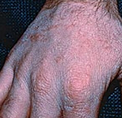 [Figure caption and citation for the preceding image starts]: Tinea manuumFrom the collection of Professor Antonella Tosti; used with permission [Citation ends].
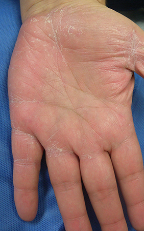
[Figure caption and citation for the preceding image starts]: Tinea pedis. Intense inflammation produces hyperpigmentation and vesicle formation. Vesiculobullous form of tinea pedisDepartment of Dermatology Medical University of South Carolina; used with permission [Citation ends]. [Figure caption and citation for the preceding image starts]: Vesiculobullous form of tinea pedis and onychomycosisFrom the collection of Professor Antonella Tosti; used with permission [Citation ends].
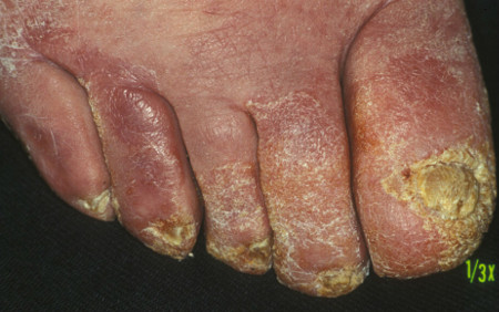
Examination of the groin
Tinea cruris affects the proximal, medial thighs. It can extend to the abdomen and buttocks. The scrotum tends to be spared. Pustules and vesicles at the active edge of the infected area, along with maceration, are present. There are also red, scaling lesions with raised borders noted in the background.
Evaluate the feet as a source of the infection.
Examination of the nail
Several clinical features suggest onychomycosis, including:
White-yellow to orange-brown patches or streaks in the nail
Onycholysis (nail plate separating from the nail bed)
Subungual debris
Hyperkeratosis (thickening) of the nail bed
Dystrophy of normal nail architecture.
Onychomycosis can be divided into four subtypes by clinical appearance:
Distal lateral subungual onychomycosis (DLSO): nails are thickened with subungual hyperkeratosis and onycholysis; discoloration ranges from white-yellow to brown
White superficial onychomycosis: confined to the toenails and presents as small, white speckled patches on the surface of the nail plate
Proximal subungual onychomycosis: presents as an area of leukonychia in the proximal nail; the nail plate surface is normal and there is no subungual hyperkeratosis
Endonyx onychomycosis: nails have a milky white colour of the nail plate, but, unlike DLSO, no evidence of subungual hyperkeratosis or onycholysis.
Patients may have a combination of the above subtypes.
Non-dermatophyte mould infection
An additional subset to consider, non-dermatophyte mould (i.e., Scopulariopsis brevicaulis, Fusarium, Aspergillus, and Acremonium species infections) onychomycosis can be difficult to diagnose given that non-dermatophyte moulds are common contaminants of the nails and of the mycology laboratory. These infections are usually very difficult to treat.[20]
[Figure caption and citation for the preceding image starts]: Distal lateral subungual onychomycosisFrom the collection of Professor Antonella Tosti; used with permission [Citation ends].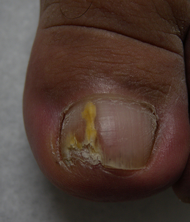
Laboratory confirmation
Method of diagnosis varies according to region. In the first instance, clinicians may use microscopy to identify fungus. Skin scrapings or nail clippings may also be sent to a mycology lab for microscopy and culture. Sending samples to mycology is recommended, if possible, for targeted treatment.[13][19][21]
The majority of nail infections start under the nail, so the subungual debris is particularly valuable. Superficial scrapings of the nail plate are only useful in cases of superficial onychomycosis, where white crumbly patches are seen on the dorsal surface of the nail, or in proximal subungual onychomycosis.
About 50% of nails in which fungus has been seen on direct microscopy fail to grow a pathogen.[22]
Fungal culture media is recommended for hair and nail infections. Fungal speciation and antifungal sensitivities may be necessary in tinea that is unresponsive to treatment, especially in tinea capitis that has been resistant to griseofulvin, or in multisite tinea cases.[21] Serial fungal cultures can aid in documentation of mycological cure, particularly for the scalp, and are recommended.[23]
Polymerase chain reaction (PCR) facilitates fast diagnosis of dermatophyte infections.[24]
Rarely, a nail biopsy is used to rule out other causes of dystrophic nails, such as psoriasis.
Confirmation of onychomycosis
Nail infection should be confirmed by pathology or mycology before starting treatment.[13]
PAS staining of nail clippings is useful to confirm diagnosis of onychomycosis.[25]
Fungal culture is useful for confirming diagnosis when long-term oral therapy is being considered, particularly after negative KOH microscopy when there is a high index of suspicion for tinea unguium. Take the sample from the under-surface of the nail, as proximally as possible.
PCR rapidly confirms the diagnosis of fungal nail infection.[26]
Confirmation of tinea capitis
Dermoscopy is a non-invasive tool for the identification of hair and scalp disorders.[27] The presence of comma and corkscrew hairs suggests a diagnosis of tinea capitis.
Fungal cultures are recommended in tinea capitis when KOH testing is non-confirmatory.[23] Sample scalp lesions by scalpel scraping, hair pluck, brush, or swab as appropriate to the lesion.[23] Fungal culture of the scalp is best accomplished by running wet cotton buds through the scalp and plating on media.
KOH testing on the hair often reveals hairs infected with sporae, whereas preparations of tinea from other sites show hyphae in the stratum corneum as common features.
Wood lamp examination (using ultraviolet light)
Rarely useful in identification of dermatophyte infection. It can only be used to diagnose tinea capitis infection by zoophilic organisms, which will fluoresce; these account for a very small percentage of diagnoses in developed countries.
Wood lamp examination may be helpful in ruling out differentials such as pityriasis versicolor or erythrasma.
Use of this content is subject to our disclaimer