Approach
An investigation, through history and physical examination, is undertaken to determine whether there are underlying causes for Eustachian tube dysfunction (ETD).
History
Patients with ETD complain of an inability to 'pop' or 'clear' the ears with changes in barometric pressure, such as when ascending or descending in a lift or an aeroplane. Some will describe this as a sense of hearing loss. A repeated history of otitis media may be also be present. Additionally, a patient may report a history of allergic rhinitis or chronic sinusitis that, when at its peak, exacerbates symptoms of fullness in the ears. A validated ETD-specific symptom score, the seven-item Eustachian Tube Dysfunction Questionnaire (ETDQ-7), is also available to quantify the severity of the condition.[28]
Patients with patulous (abnormally patent) Eustachian tubes will describe a sense of autophony (hearing one's voice or breath in the ear), which resolves with supine position or leaning the head forwards. Patulous Eustachian tubes are often associated with significant recent weight loss.
Physical examination
Patients with ETD may have an entirely normal head and neck examination. Pertinent findings include: a retracted or hypomobile tympanic membrane, particularly in the weak pars flaccida area; a serous otitis media; an inflamed or oedematous Eustachian tube orifice on endoscopic examination; adenoid hypertrophy; or neoplasm filling the nasopharynx.
Initial testing
Tympanometry, measuring the compliance of the tympanic membrane, reveals a type B or type C tympanogram. A normal (type A) tympanogram shows normal middle ear pressure and a normally functioning Eustachian tube. A type B tympanogram has a flat compliance curve, demonstrating no movement of the tympanic membrane. A type C tympanogram shows a shift of the compliance curve with negative pressure. Quantitative values of tympanometric peak pressure may provide more specific information than morphologic categorisation. [Figure caption and citation for the preceding image starts]: Normal (type A) tympanogramFrom the collection of Erica R. Thaler; used with permission [Citation ends]. [Figure caption and citation for the preceding image starts]: Type B tympanogram; flat compliance curve demonstrates no movement of the tympanic membraneFrom the collection of Erica R. Thaler; used with permission [Citation ends].
 [Figure caption and citation for the preceding image starts]: Type C tympanogram, demonstrating a malfunctioning Eustachian tubeFrom the collection of Erica R. Thaler; used with permission [Citation ends].
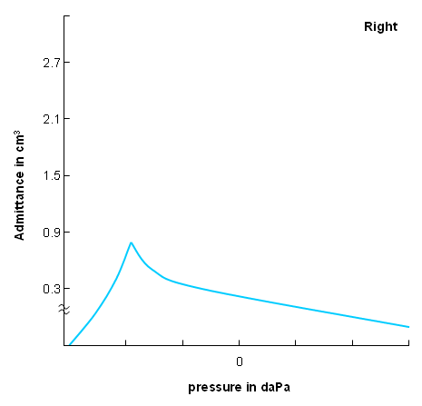
Nasal endoscopy is a routine part of an otolaryngologist's assessment of a patient with ETD, and may show oedema or obstruction of the Eustachian tube orifice.[Figure caption and citation for the preceding image starts]: A normal left Eustachian tube as seen endoscopically from the posterior nasal cavity.From the collection of Edward D. McCoul; used with permission [Citation ends].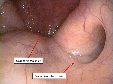
Subsequent testing
In adults with unilateral serous otitis, a nasopharyngoscopy and/or CT scan should be done to look for any mass in the nasopharynx or infratemporal fossa.
Use of this content is subject to our disclaimer