Images and videos
Images


Coxiella burnetii infection
Natural history of Coxiella burnetii and serological/PCR results. Ac+: positive for antibodies to C burnetii, Ac++: strongly positive for antibodies to C burnetii, Ac-: negative for antibodies to C burnetii, PCR+: positive PCR for C burnetii, PCR++: strongly positive PCR for C burnetii, PCR-: negative PCR for C burnetii
Eldin C, et al. Clin Microbiol Rev 2017; used with permission
See this image in context in the following section/s:

Coxiella burnetii infection
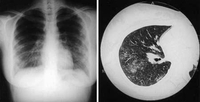
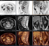
Coxiella burnetii pneumonia. CXR and CT scan from a 21-year-old woman with Coxiella burnetii pneumonia; CXR shows multiple areas of soft consolidation in the middle-to-lower lung fields bilaterally; CT scan shows poorly defined centrilobular nodules and air space consolidation
Okimoto N, et al. Respirology. 2004;9:278-282; used with permission of John Wiley & Sons Ltd
See this image in context in the following section/s:
Coxiella burnetii infection
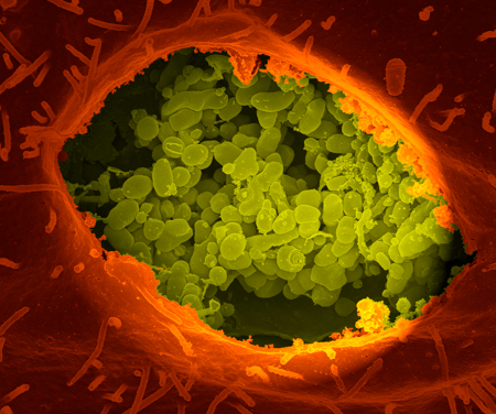
Coxiella burnetii in the vacuole. A dry fracture of a vero cell exposing the contents of a vacuole where Coxiella burnetii are growing
National Institute of Allergy and Infectious Diseases (NIAID); National Institutes of Health (NIH)
See this image in context in the following section/s:

Coxiella burnetii infection
Coxiella burnetii lung pseudotumour: fluorescence in situ hybridisation (FISH). Red: immunofluorescence (IF). Green: FISH with a specific 16S rRNA probe. Yellow: co-localisation of IF and FISH confirms the presence of bacteria in the cytoplasm of 2 cells in a lung pseudotumour
Gilles Audoly, Institut Hospitalo-Universitaire Méditerranée Infection
See this image in context in the following section/s:

Coxiella burnetii infection
Algorithm for the diagnosis and management of C burnetiiinfection. TTE: transthoracic echocardiography; IgG aCL: IgG anticardiolipin antibodies; 18 F-FDG PET/CT: 18F-fluorodeoxyglucose PET combined with CT
Eldin C, et al. Clin Microbiol Rev 2017; used with permission
See this image in context in the following section/s:

Coxiella burnetii infection
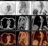
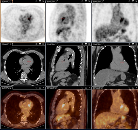
Q fever aortic mycotic thoracic aneurysm diagnosed at PET scan: 18F-fluorodeoxyglucose PET/CT. In this asymptomatic patient with heart valve history with elevated serology, the PET scan diagnosed an aortic endocarditis on native valve with thoracic and lumbar aortic mycotic aneurysms
Institut Hospitalo-Universitaire Méditerranée Infection (patient consent obtained)
See this image in context in the following section/s:
Coxiella burnetii infection
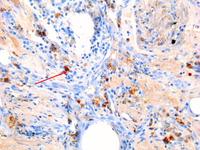
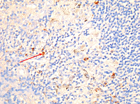
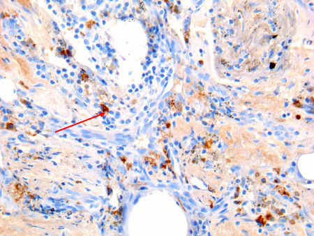
Coxiella burnetii chronic lymphadenitis: immunohistochemistry. Note the isolated infected cell (monocytes/macrophages) in the lymph node. Brown coloration identifies bacteria in monocytes/macrophages
Hubert Lepidi, Institut Hospitalo-Universitaire Méditerranée Infection
See this image in context in the following section/s:
Coxiella burnetii infection
Q fever endocarditis diagnosed at PET scan: 18F-fluorodeoxyglucose PET/CT. In this asymptomatic patient with heart valve history with elevated serology, the PET scan diagnosed an aortic endocarditis on native valve with thoracic and lumbar aortic mycotic aneurysms
Institut Hospitalo-Universitaire Méditerranée Infection (patient consent obtained)
See this image in context in the following section/s:
Coxiella burnetii infection
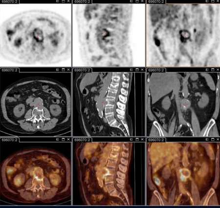
Q fever aortic mycotic lumbar aneurysm diagnosed at PET scan: 18F-fluorodeoxyglucose PET/CT. In this asymptomatic patient with heart valve history with elevated serology, the PET scan diagnosed an aortic endocarditis on native valve with thoracic and lumbar aortic mycotic aneurysms
Institut Hospitalo-Universitaire Méditerranée Infection (patient consent obtained)
See this image in context in the following section/s:
Coxiella burnetii infection
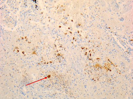
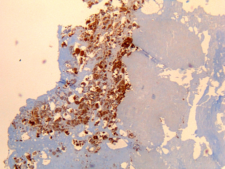
Coxiella burnetii lung fibrosis: immunohistochemistry; brown coloration identifies bacteria in monocytes/macrophages
Hubert Lepidi, Institut Hospitalo-Universitaire Méditerranée Infection
See this image in context in the following section/s:
Coxiella burnetii infection
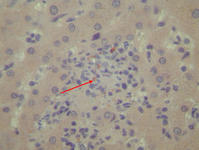
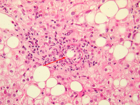
Liver 'doughnut granuloma' characteristic of acute Coxiella burnetii hepatitis. Note the specific granuloma morphology as a doughnut. No bacteria may be seen in this granuloma
Hubert Lepidi, Institut Hospitalo-Universitaire Méditerranée Infection
See this image in context in the following section/s:
Coxiella burnetii infection
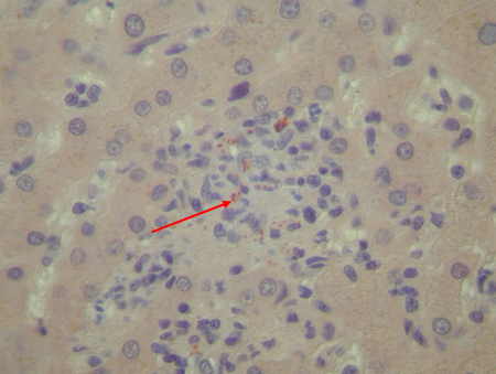
Coxiella burnetii endocarditis: immunohistochemistry. Note the low level of inflammation. Brown coloration identifies bacteria in monocytes/macrophages inside. Vegetation is usually lacking
Hubert Lepidi, Institut Hospitalo-Universitaire Méditerranée Infection
See this image in context in the following section/s:
Coxiella burnetii infection
Coxiella burnetii osteitis: immunohistochemistry: brown coloration identifies bacteria in monocytes/macrophages
Hubert Lepidi, Institut Hospitalo-Universitaire Méditerranée Infection
See this image in context in the following section/s:
Coxiella burnetii infection
Coxiella burnetii chronic hepatitis of a patient with endocarditis: immunohiostochemistry. Note the absence of doughnut granuloma seen in acute Q fever. Brown coloration identifies bacteria in monocytes/macrophages
Hubert Lepidi, Institut Hospitalo-Universitaire Méditerranée Infection
See this image in context in the following section/s:
Use of this content is subject to our disclaimer