Investigations
1st investigations to order
clinical diagnosis
Test
Diagnosis of oral mucositis (OM) is typically based upon the clinical history and physical examination.[1][2]
Common sites include the buccal mucosa, lip mucosa, ventral tongue, and soft palate.[Figure caption and citation for the preceding image starts]: Mucositis: 'buccal' mucosaFrom the teaching collection of Rajesh V. Lalla, DDS, PhD, CCRP, DABOM; used with permission [Citation ends].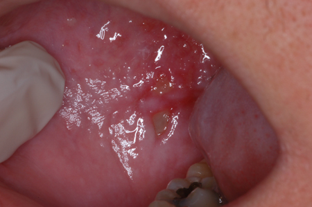
[Figure caption and citation for the preceding image starts]: Mucositis: dorsolateral tongueFrom the teaching collection of Rajesh V. Lalla, DDS, PhD, CCRP, DABOM; used with permission [Citation ends].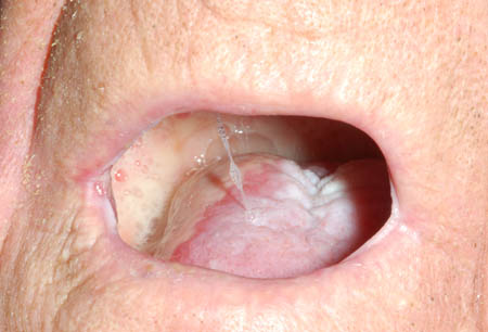
The most important diagnostic factor is a history of chemotherapy and/or radiotherapy (to the oral cavity) for the treatment of cancer.
Usually, no tests are necessary; however, in certain circumstances, investigations testing may be warranted to rule out other conditions that may mimic or complicate mucositis.
Result
clinical examination: features of OM range from erythema to patchy or confluent ulceration (often with a superficial pseudomembranous membrane) or, rarely, overt necrosis; lesions may be unilateral or bilateral, and are typically limited to non-keratinised oral mucosa
Investigations to consider
FBC with differential
Test
Order immediately in any patient where febrile neutropenia is suspected.
See Febrile neutropenia.
Result
variable; may show leukocytosis and/or neutropenia; WBC and neutrophil (ANC) counts should be interpreted in the context of any associated malignancy; however, in the presence of fever, an ANC <500 cells/microlitre, or an ANC <1000 cells/microlitre with a projected ANC of <500 cells/microlitre, suggests febrile neutropenia
blood cultures
Test
Order immediately in any patient where febrile neutropenia is suspected.
See Febrile neutropenia.
Result
may be positive for a pathogen (particularly in the presence of neutropenia)
superficial smear of lesion for microscopy
Test
Consider if secondary infection with Candida is suspected, or an alternative diagnosis to oral mucositis is more likely.
In pseudomembranous candidiasis, Candida hyphae are numerous; in erythematous candidiasis, hyphae are few in number and additional testing with culture may be necessary.
Candida albicans can also be a normal inhabitant of the oral cavity. Therefore, results need to be interpreted according to the number of colony-forming units and taking clinical findings into consideration. Other species (Candida glabrata and Candida tropicalis) may also be found.[39]
Result
variable; may show Candida hyphae
fungal culture
Test
Consider if secondary infection with Candida is suspected, or an alternative diagnosis to oral mucositis is more likely.
In pseudomembranous candidiasis, Candidahyphae are numerous and may be confirmed by microscopic examination of a superficial smear; in erythematous candidiasis, hyphae are few in number and additional testing with culture may be necessary.
Candida albicans can also be a normal inhabitant of the oral cavity. Therefore, results of the fungal culture need to be interpreted according to the number of colony-forming units and taking clinical findings into consideration. Other species (Candida glabrata and Candida tropicalis) may also be found.[39]
Result
variable; may be positive for Candida
viral culture or polymerase chain reaction (PCR)
Test
Consider if secondary infection with herpes simplex virus (HSV) is suspected, or an alternative diagnosis to oral mucositis is more likely.
PCR has a higher sensitivity than viral culture, but may not be available in some locations.[43]
Result
variable; may be positive for HSV infection
Use of this content is subject to our disclaimer