Aetiology
The aetiology of nasal polyps is not fully understood. They seem to arise due to chronic mucosal inflammation. Chronic rhinosinusitis with nasal polyps (CRSwNP) is closely associated with adult-onset, eosinophilic asthma.[3] There is likely to be a shared aetiology as well as shared pathology, in such patients, but the precipitating factors are unknown.
Genetic associations have been reported in some cases of CRSwNP, but the evidence is limited.[8][9][10] Population studies have demonstrated a four- to fivefold increased risk of CRSwNP in people who have a first-degree relative with CRSwNP.[10][11] Specific risk genes are not yet well characterised.[12] The role of environmental factors is unclear.[2]
People with aspirin- and non-steroidal anti-inflammatory drug (NSAID)-exacerbated respiratory disease have a higher incidence of nasal polyps (estimates range from 7% to 26%).[3] The combination of CRSwNP, asthma, and aspirin/NSAID hypersensitivity is commonly referred to as Samter's triad.[13]
Worldwide, allergic fungal rhinosinusitis may account for 5% to 10% of CRSwNP cases.[14][15] Here, it is assumed that an intense, IgE-driven, eosinophilic inflammatory response occurs to fungal hyphae that are able to colonise the nasal sinuses, such as Aspergillus species.[16]
Pathophysiology
There is diversity in cytokine levels, eosinophilic/neutrophilic patterns, and IgE expression among patients with CRSwNP from Europe, Asia, and Oceania.[17] Histologically, nasal polyps from patients of European descent consist of loose connective tissue, oedema, inflammatory cells, mucous glands, and capillaries. They are predominantly covered with respiratory pseudostratified epithelium with ciliated and goblet cells. The stromal inflammatory cells of nasal polyps from patients of European descent are mainly characterised by eosinophils, although neutrophils, mast cells, plasma cells, lymphocytes, monocytes, and fibroblasts are also seen.[1][2][Figure caption and citation for the preceding image starts]: Grade 3 nasal polypsFrom the collection of Dr Richard Hewitt [Citation ends].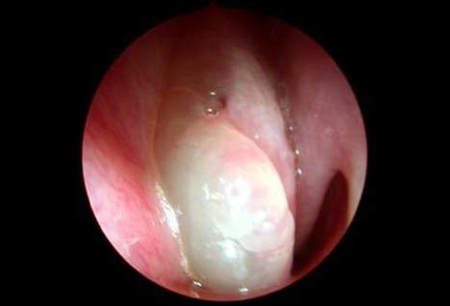
Most nasal polyps from European populations are associated with profound local Th2-type inflammation and are often accompanied by a mild peripheral blood eosinophilia, particularly if accompanied by asthma. Nasal polyp tissue from patients of European descent is rich in interleukin-5, the key cytokine for eosinophil recruitment (from the bone marrow) and survival. By contrast, nasal polyp tissue from patients of Chinese descent is rich in neutrophils but low in eosinophils, with a Th1/Th17-type inflammatory profile.[17] Polyp tissue from patients of European descent may also express high levels of IgE antibodies, while some studies have shown IgE expression to be low in polyp tissue from patients of Chinese descent.[17][18]
Atopy - the presence of positive skin-prick tests/serum IgE antibodies to common aeroallergens - is not clearly related to CRSwNP; the IgE produced within polyp tissue may be relatively non-allergen-specific. Evidence for the pathological importance of interleukin-5 and IgE in the pathophysiology of CRSwNP comes from studies of monoclonal antibody drugs that target these proteins.[19][20][21][22]
Classification
Anatomical and histological classification
Nasal polyps may be classified according to anatomy, histology and, to some extent, in relation to underlying disease.
Most polyps are ethmoidal - arising from the ethmoid sinus and extending into the nasal cavity. Ethmoidal polyps are often multiple, with an appearance resembling a bunch of grapes.[1] They are usually bilateral. A minority of polyps are antrochoanal - arising in the maxillary sinus and extending toward the pharynx. They are typically larger and single, and more common in children than adults.
A minority of polyps occur on a background of other underlying diseases beyond simple chronic rhinosinusitis with nasal polyps, including eosinophilic polyangiitis (formerly Churg-Strauss syndrome), cystic fibrosis, and allergic fungal rhinosinusitis.
Use of this content is subject to our disclaimer