Images and videos
Images


Assessment of liver dysfunction
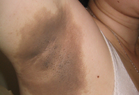
Acanthosis nigricans involving the axilla of an obese white woman
From the collection of Melvin Chiu, MD, UCLA
See this image in context in the following section/s:
Assessment of liver dysfunction
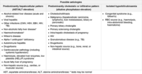
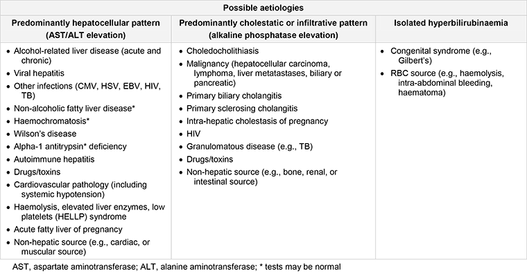
Possible diagnosis for underlying cause of different patterns of liver test abnormalities
Created by the BMJ Group
See this image in context in the following section/s:
Assessment of liver dysfunction
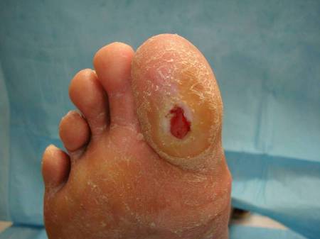
Plantar ulcer in a patient with type 1 diabetes
From the collection of Rodica Pop-Busui, MD, PhD, University of Michigan, MI
See this image in context in the following section/s:

Assessment of liver dysfunction
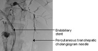
Typical ERCP findings in a patient with primary sclerosing cholangitis: multi-focal strictures of the intra- and extrahepatic bile ducts
From the collection of Kris V. Kowdley, MD, University of Washington, WA
See this image in context in the following section/s:
Assessment of liver dysfunction
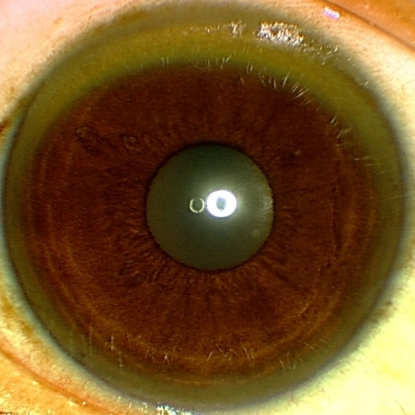
Eye demonstrating Kayser-Fleischer ring
Adapted from BMJ (2009), used with permission; copyright 2009 by the BMJ Publishing Group
See this image in context in the following section/s:

Assessment of liver dysfunction
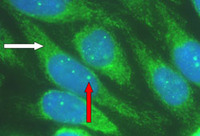
Characteristic auto-antibody patterns in primary biliary cholangitis. White arrow: anti-mitochondrial staining; red arrow: multiple nuclear dot ANA staining
From the collection of David E.J. Jones, BA, BM, BCh, PhD, FRCP, University of Newcastle, UK
See this image in context in the following section/s:
Assessment of liver dysfunction
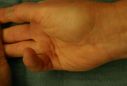
Dupuytren's contracture
From the collection of Craig M. Rodner, MD, University of Connecticut Health Center/New England Musculoskeletal Institute, CT
See this image in context in the following section/s:
Use of this content is subject to our disclaimer