Images and videos
Images


Bartholin's cyst
Insertion of a Word catheter to treat a Bartholin’s cyst. The site is cleaned and anaesthetised, and then a small stab incision (3 to 4 mm) is made into the cyst/abscess cavity (parallel to the hymen ring). The Word catheter is introduced into the cyst/abscess cavity after the contents have drained. The balloon is filled with sterile saline and a suture is tied around the catheter to prevent leaking or deflation. The catheter end is then tucked into the vagina.
From the personal collection of Colleen Kennedy Stockdale; used with permission
See this image in context in the following section/s:
Bartholin's cyst
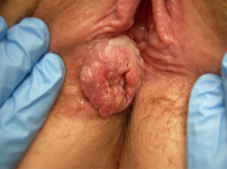
Squamous cell cancer
From the personal collection of Colleen Kennedy Stockdale
See this image in context in the following section/s:
Bartholin's cyst
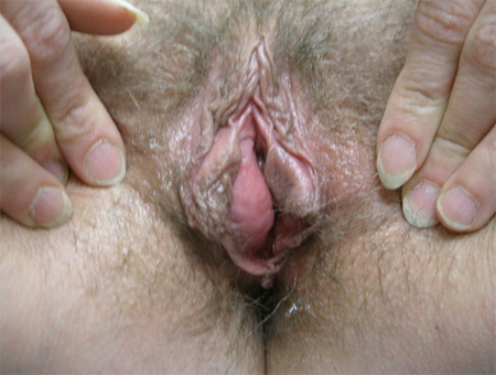
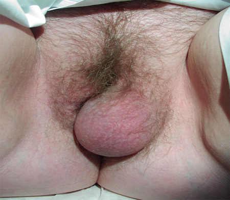
Bartholin's duct cyst at posterior labia minora, which crosses central portion of cyst with half of the enlargement medial and the other half lateral to this line
From the personal collection of Colleen Kennedy Stockdale
See this image in context in the following section/s:
Bartholin's cyst
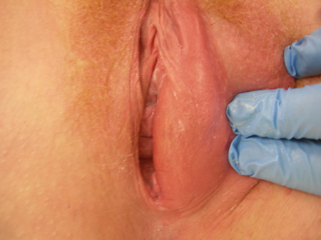
Bartholin's abscess: erythema, oedema, with cyst abscess posterior to labia minora
From the personal collection of Colleen Kennedy Stockdale
See this image in context in the following section/s:
Bartholin's cyst
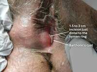
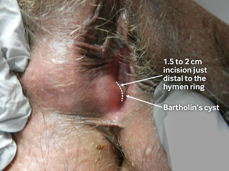
Marsupialisation of a Bartholin’s cyst. The site is cleaned and anaesthetised, and then a 1.5 to 2 cm incision is made just distal to the hymen ring within the introitus into the region of the normal duct opening. The cyst/abscess cavity is irrigated and loculations are broken down if necessary. The incised cyst/abscess wall is then approximated to the edge of the vestibular skin.
From the personal collection of Colleen Kennedy Stockdale; used with permission.
See this image in context in the following section/s:

Bartholin's cyst
Bartholin's cyst
From the personal collection of Colleen Kennedy Stockdale
See this image in context in the following section/s:
Bartholin's cyst
Vulval lipoma
From the personal collection of Colleen Kennedy Stockdale
See this image in context in the following section/s:
Use of this content is subject to our disclaimer