Approach
The diagnosis of Bartholin's cysts and abscesses is clinical and readily made on visual inspection of the vulva. Further investigations are not required to make the diagnosis.
History
They may be asymptomatic and identified by chance during a pelvic exam. Most Bartholin's cysts occur in reproductive-age women between 20 and 50 years.[10] Symptoms may include pain on sitting or walking, or during sexual intercourse.[17][18] The size of the cyst depends on accumulation of gland secretions, and is exemplified by rapid enlargement during sexual activity and shrinkage or stability of cyst size in women with diminished sexual activity. Patients may also complain of a feeling of fullness or pressure, particularly with increasing cyst size.[12] Most abscesses usually develop rapidly and are associated with severe pain. About one quarter of women with Bartholin's abscess have fever.[15]
Physical examination
The classic, medially protruding cystic structure at the inferior aspect of the labia majora (in the 5 or 7 o'clock position) crossed by the labium minus is typical. [Figure caption and citation for the preceding image starts]: Bartholin's cystFrom the personal collection of Colleen Kennedy Stockdale [Citation ends].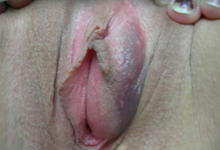 [Figure caption and citation for the preceding image starts]: Bartholin's duct cyst at posterior labia minora, which crosses central portion of cyst with half of the enlargement medial and the other half lateral to this lineFrom the personal collection of Colleen Kennedy Stockdale [Citation ends].
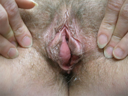 Occasionally, cysts dissect anterolaterally into the labium majus.[2] When infection is present or an abscess has formed, the mass is usually tender, and there may be induration, erythema, point discharge, or systemic fever. [Figure caption and citation for the preceding image starts]: Bartholin's abscess: erythema, oedema, with cyst abscess posterior to labia minoraFrom the personal collection of Colleen Kennedy Stockdale [Citation ends].
Exclusion of other causes
Any vulval mass that is not classically crossed by the labium minus at the inferior aspect of the labia is unlikely to be a Bartholin's cyst.
Most other causes of vulval swellings, such as epidermal inclusion cysts, mucous cysts, or cysts of the canal of Nuck, are located in the labia majora and are not transected by the labium minus.
Skene's ducts are located adjacent to the urethral meatus rather than the posterior vestibules. While treatment may also be warranted for other vulval cysts, the approach may be different, according to the aetiology.
Although Bartholin's gland cancer is exceedingly rare (incidence <1 in 500,000 women), it should be considered in women presenting with an irregular/firm Bartholin's process.[6] The classic presentation of Bartholin's gland cancer is an irregular, nodular mass in the region of the duct/gland.[2] The incidence is highest among women in their 60s but should be considered in all women with nodularity, irregularity, or persistent induration.[19][Figure caption and citation for the preceding image starts]: Squamous cell cancerFrom the personal collection of Colleen Kennedy Stockdale [Citation ends].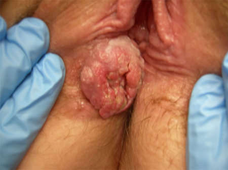
Investigations
Routine blood tests are not required.
Microbiological culture and sensitivities of abscess material are not necessary to diagnose a Bartholin's abscess. They may be helpful in identifying the predominant organism and targeting antimicrobial treatment when resolution is incomplete after incision and drainage. More than 70% of cultures from Bartholin's cysts and about 33% of cultures from Bartholin's abscesses are sterile.[13][14]
A lesion biopsy should be performed if malignancy is suspected based on clinical appearance. It should also be considered in women >40 years of age with a Bartholin's cyst or abscess, given the increased incidence of Bartholin's gland cancer among menopausal women.
Use of this content is subject to our disclaimer