Images and videos
Images


Retinoblastoma
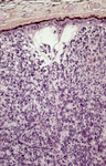
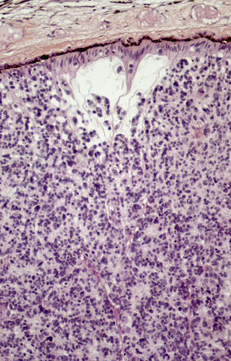
Histopathology of retinoblastoma. This image demonstrates the classical features of retinoblastoma, including densely packed, small, round tumor cells with hyperchromatic nuclei and scant cytoplasm, arranged in sheets. The absence of Flexner-Wintersteiner rosettes in this specimen does not preclude the diagnosis, as their presence is not obligatory. These histopathological features are typical of this aggressive retinal tumor and provide critical information for diagnosis, staging, and prognosis
PR J. L. Kemeny, ISM/Science Photo Library; used with permission
See this image in context in the following section/s:
Retinoblastoma
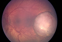
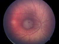
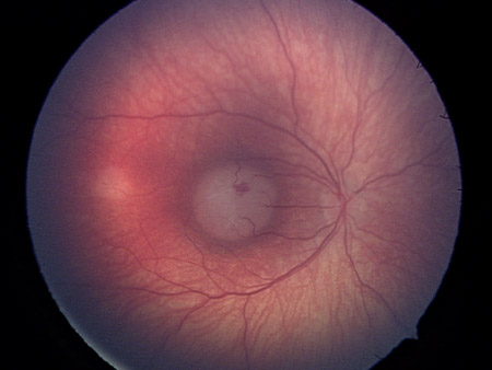
Macular retinoblastoma in the right eye
Personal collection of Dr Timothy Murray
See this image in context in the following section/s:

Retinoblastoma
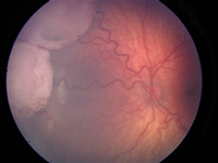
Vitreous seeding associated with retinoblastoma
Personal collection of Dr Timothy Murray
See this image in context in the following section/s:
Retinoblastoma
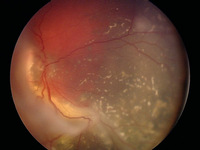
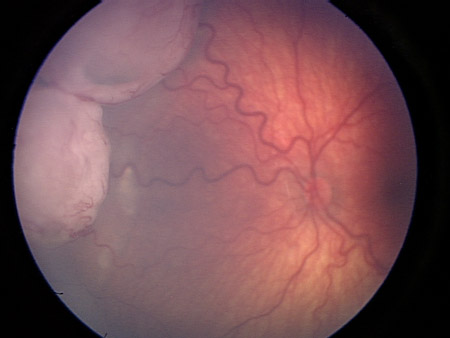
Two large retinoblastoma foci in the left eye; note the associated subretinal seeding
Personal collection of Dr Timothy Murray
See this image in context in the following section/s:

Retinoblastoma
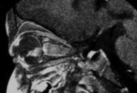
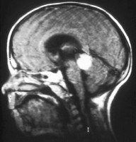
MRI pattern of retinoblastoma with optic nerve involvement (sagittal enhanced T1-weighted sequence)
Aerts I, et al. Orphanet J Rare Dis 2006 Aug 25; 1: 31; licensed under CC BY 2.0
See this image in context in the following section/s:
Retinoblastoma
Aspect of trilateral retinoblastoma (MRI)
Aerts I, et al. Orphanet J Rare Dis 2006 Aug 25; 1: 31; licensed under CC BY 2.0
See this image in context in the following section/s:
Retinoblastoma
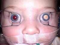
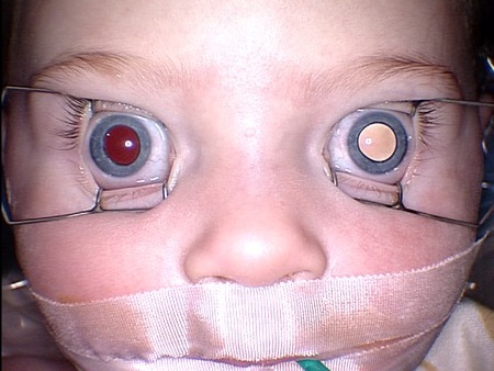
Leukocoria (white pupillary light reflex) in the left eye of a patient with unilateral retinoblastoma
Personal collection of Dr Timothy Murray
See this image in context in the following section/s:
Retinoblastoma
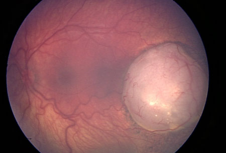
Large retinoblastoma focus in the left eye
Personal collection of Dr Timothy Murray
See this image in context in the following section/s:
Retinoblastoma
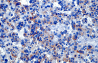
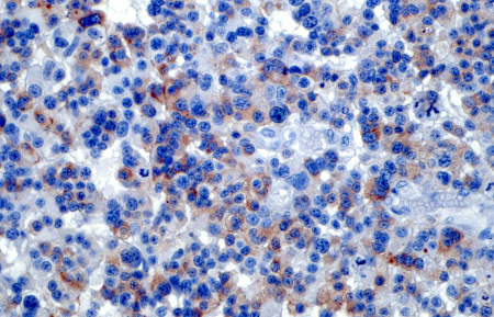
Light micrograph of a retinal section from a patient with retinoblastoma, a rare form of intraocular cancer. The tumor shows disrupted retinal architecture and infiltrative growth of atypical cells. Immunohistochemistry with anti-rhodopsin antibodies highlights areas of preserved photoreceptor differentiation within the retinal tissue. This staining aids in identifying residual retinal layers amidst the malignant cellular proliferation
PR J. L. Kemeny, ISM/Science Photo Library; used with permission
See this image in context in the following section/s:
Retinoblastoma
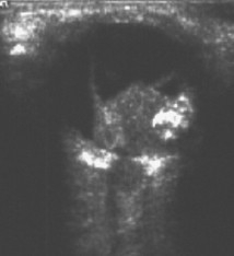
Ultrasound of retinoblastoma
Aerts I, et al. Orphanet J Rare Dis 2006 Aug 25; 1: 31; licensed under CC BY 2.0
See this image in context in the following section/s:
Use of this content is subject to our disclaimer