Approach
ABC (airway, breathing, circulation) should be implemented as required in unstable patients. In stable or stabilised patients, removal of a foreign body can be achieved after initial investigation by various methods, the choice of which will depend on the site of the foreign body, risk, and haemodynamic stability of the patient.
Management of a foreign body should be handled on a case by case basis. The likelihood that the foreign body will pass spontaneously and be evacuated in the stool, without causing damage while passing through the alimentary canal, depends upon its size, shape, composition, and whether the patient has anatomical abnormalities that could predispose to retention.[Figure caption and citation for the preceding image starts]: Diagnostic and treatment algorithm for foreign body ingestionFrom the collection of Juan Carlos Munoz [Citation ends].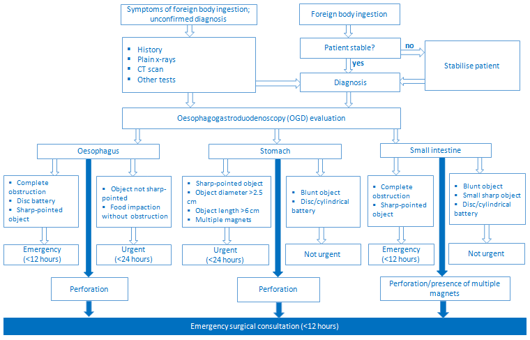
Initial management of an unstable patient
Patients should be considered unstable if they present with airway compromise, drooling, inability to tolerate fluids, evidence of sepsis, perforation, or active bleeding. The initial diagnostic evaluation involves an assessment of haemodynamic instability and resuscitation efforts. If necessary, the ABCs (airway, breathing, and circulation) should be implemented.
Elective endotracheal intubation should be considered in patients with respiratory compromise or distress, mental status alteration, or active bleeding. Elective endotracheal intubation will protect the airway and facilitate endoscopy. Bilateral peripheral or central intravenous access should be obtained for adequate fluid resuscitation. Resuscitative measures as appropriate, including volume resuscitation (crystalloid infusion), oxygen (if O₂ saturation is <90%), blood product replacement with packed red blood cell transfusion for ongoing bleeding (haemoglobin <70 g/L [<7 g/dL] or <80 g/L [<8 g/dL] with cardiac issues), and correction of coagulopathy should be instituted.
As soon as the patient is stable, a complete history and physical examination (from mouth to anus) can be performed with relevant interventions applied as required according to the stable patient group. Perforation may be suspected and confirmed by the presence of subcutaneous emphysema, pneumomediastinum, pneumothorax, or pleural effusion on posteroanterior and lateral x-rays of the neck and chest, computed tomography scan of neck and chest, or gastrografin swallow. In patients with signs or symptoms of septicaemia or elevated white blood cell count at presentation, intravenous broad-spectrum antibiotics should be started.
Emergency surgery is mandatory in patients with perforation due to obstruction, with the goal of cleaning out intra-abdominal contamination via irrigation, resecting the area of perforation, and addressing the underlying problem.
Ingestion of multiple magnets is serious and requires special attention and follow-up.[43] Magnets in adjacent intestinal loops may attract each other with significant force, causing intestinal obstruction, pressure necrosis of the intestinal wall leading to a perforation, or fistula formation.[3][19] Early endoscopic or surgical intervention is crucial, especially if the patient is symptomatic or sequential abdominal radiology films suggest no change in the position of the magnets.[3][19][54]
Urgent versus emergency foreign body removal
Urgent endoscopy (within 24 hours) should be considered in the following situations: food or foreign body in the oesophagus that does not pass spontaneously but is not causing a complete oesophageal obstruction; irregular objects with sharp or pointed edges seen in the oesophagus or stomach; or a large object (>6 cm long or >2.5 cm in diameter) in the stomach. For those objects seen or impacted in the oropharynx, consider an ear, nose, and throat evaluation for possible laryngoscopy and extraction. Surgical evaluation should be considered if endoscopic extraction of the foreign body fails or is contraindicated, or a large object (>10 cm) is seen in the rectum. The European Society of Gastrointestinal Endoscopy recommends emergency (within 2 to 6 hours) therapeutic oesophagogastroduodenoscopy in the following situations: foreign bodies inducing complete oesophageal obstruction, sharp-pointed objects, or batteries in the oesophagus.[34]
Special considerations apply to small disc or button battery ingestion. Although most button batteries pass through the gut uneventfully and are eliminated in the stool, batteries that lodge and become impacted in the oesophagus can cause life-threatening complications within 2 hours.[56] Complications (which may be delayed up to 18 days after battery removal) include oesophageal perforation, tracheo-oesophageal fistula, aorto-oesophageal fistula with subsequent exsanguination from arterial fistulisation, electrical discharge, chemical injury with subsequent oesophageal strictures, retropharyngeal abscess, mediastinitis, vocal cord paralysis, and tracheal stenosis. These complications should be monitored vigorously after the battery is removed, and patients advised to report symptoms urgently.
Injury is thought to be secondary to electrochemical burns from electrical discharge. Chemical burns from electrolyte alkali leakage and pressure necrosis may also contribute.[16] The 3-volt, 20 mm-diameter (CR/BR type) lithium button battery is particularly prone to lodging in the oesophagus, especially in children. An immediate x-ray should be obtained to determine battery location and diameter, and the battery's chemical composition and type should be determined where possible from packaging or a matching battery. Oesophageal impaction of a battery requires emergency endoscopic or surgical removal.[43]
For patients without oesophageal impaction, conservative intervention is recommended in the absence of symptoms and signs of injury.[57][58] Retrieval is indicated only if two or more magnets are ingested at the same time, symptoms develop, or a large diameter battery (>15 mm) fails to pass the pylorus in 4 days. All other batteries beyond the oesophagus may be managed at home with regular diet and activity. If passage is not documented within 10 to 14 days, repeat radiographs can be performed to confirm passage of the battery.[56]
Ingestion of multiple magnets constitutes a unique problem. Early endoscopic removal should be the treatment of choice when objects are lodged in the oesophagus or stomach. If they have passed beyond the pylorus the retraction could be more difficult, if not impossible. An emergency surgical consultation should be made.[19]
Removal of foreign body in a stable patient
Spontaneous passage with no intervention (watchful waiting)
The decision to 'wait and see' will depend on several factors: age of the patient, physical characteristics of the object, location, and symptoms of obstruction. This option can be considered in children with single coin ingestion and in adults with foreign objects that are distal to the oesophagus. These patients can be observed (stools should be searched for the object or serial x-rays performed) to determine whether the object will advance to the rectum.
Fibre-optic nasopharyngoscopy
Ear, nose, and throat evaluation should be considered for those patients in whom an object is in the oropharynx that cannot be seen during the physical examination.
Removal of foreign bodies can be accomplished using a tongue depressor and laryngoscopy.[13][59]
Fibre-optic oesophagogastroduodenoscopy, flexible sigmoidoscopy, and colonoscopy
The forward-viewing flexible endoscope has become the instrument of choice in managing most impacted foreign bodies. Emergency endoscopy is indicated for patients whose airway is compromised, those who show signs of complications, or if a battery is impacted in the oesophagus. Endoscopy is required for foreign bodies that are non-progressive, sharp, or non-radiopaque; batteries; multiple foreign bodies; foreign bodies in the stomach >6 cm long or >2.5 cm in diameter; or oddly shaped foreign bodies such as open safety pins. 'Side-viewing' flexible endoscopy has been used to diagnose and manage foreign bodies in the common bile duct.[60] Enteroscopy (single or double balloon) may allow the removal of foreign bodies in the small intestine, when indicated.[45]
The following tools can be used to assist in the endoscopic removal of foreign bodies: forceps, snares, Roth retrieval net, Dormia baskets, balloons, overtubes, and latex protector hood. Despite potential technical challenges, endoscopy has made the removal of foreign bodies possible without surgical intervention (which is only needed in approximately 1% of cases); furthermore, it has emerged as one of the safest methods of managing foreign bodies in the gastrointestinal (GI) tract.[59][61][62]
For encased foreign bodies, extraction may be difficult or impossible. In this case it may be useful to postpone extraction and initiate a short course of an intravenous corticosteroid for 12 to 24 hours. This can reduce inflammation and facilitate foreign body extraction at a later time.[21]
Foley catheter removal
Foley catheter removal is another widely used technique for the removal of single, smooth, blunt, radiopaque foreign bodies from the oesophagus or rectum. This technique should be attempted only by those familiar with its use. The removal is performed under fluoroscopy guidance with immediate availability of equipment and personnel with expertise in emergency airway management. Endoscopic removal by a gastroenterologist is often a much safer option.
Foley catheter oesophageal extraction is contraindicated in the following situations: patients with an impacted foreign body that has been present longer than 72 hours, a history of oesophageal surgery, or patients who are unstable or uncooperative.
The procedure consists of placing the patient in a head-down position and a Foley catheter being passed through the mouth until it is placed behind/distal to the object. The balloon is then inflated; finally, the catheter and the object are pulled out as a unit. A success rate of 85% to 100% has been reported.[63][64]
Complications related to this procedure include epistaxis, dislodgement of the object causing laryngospasm, hypoxia, and aspiration.[63][64][65]
Endoscopic removal: adverse events
Adverse events attributable to endoscopic removal of foreign bodies are rare. The most commonly reported events are superficial mucosal lacerations (≤2%), GI bleeding (≤1%), and perforation (≤0.8%). The risk of aspiration during foreign body removal is rare, and can be minimised by using an oesophageal overtube and/or endotracheal intubation to protect the airway.[66]
Surgery
For failed extraction or objects of >10 cm in the rectum, surgical intervention may be needed. Laparoscopy can be a valuable tool in the management of GI foreign bodies, particularly when an object is not progressing or endoscopy is unsuccessful or dangerous. Other indications for surgical intervention include perforation and complications that cannot be resolved endoscopically, such as excessive bleeding.
The ingestion of multiple magnets is a special case, as they may cause adjacent intestinal loops to be forcefully attracted to each other and produce obstruction, pressure necrosis of the intestinal wall leading to a perforation, or fistula formation.[3][19] Urgent surgical intervention, such as explorative laparotomy to remove the objects and repair any damage, is vital, especially if the patient is symptomatic or sequential abdominal radiology films suggest no change in the position of the magnets.[3][19][54]
Most rectal foreign bodies can be removed transanally. After radiological confirmation of the presence, size, and location of the object, the patient is sedated (often with an intravenous sedative and/or perianal nerve block) and then placed in the left lateral or lithotomy position. Various retractors and clamps commonly used in routine anorectal surgery have been used successfully to grasp and remove objects from this area.[67]
Adjunctive treatments in stable and unstable patients
Antiemetics are useful for controlling nausea and vomiting. Commonly prescribed agents include promethazine, ondansetron, or prochlorperazine. In those with oesophageal foreign bodies, glucagon can be given intravenously to attempt spontaneous passage of an impacted foreign body. It should be infused slowly to prevent nausea and vomiting.[68][69][70][71] If there is no response, it can be repeated after 20 to 30 minutes.[69]
Intravenous antibiotics should be considered in certain cases with infective complications, after blood cultures have been taken. In cases of complete obstruction, a short course (12-24 hours) of an intravenous corticosteroid may reduce inflammation and facilitate extraction of the object at a later time.[21]
Caution during foreign body obstruction management
In the management of patients with foreign body ingestion, the following advice should be observed:
Patients' symptoms of foreign body obstruction should not be ignored.
No attempt should be made to remove hard, sharp, or large objects without using the proper equipment.
Oral enzymes such as trypsin, papain, or chymotrypsin should not be used to remove food impacted in the oesophagus. In general, this is not effective, has been associated with hypernatraemia, and carries the risk of enzyme-induced oesophageal perforation.
Glucagon is contraindicated in patients with a phaeochromocytoma or insulinoma.
Enemas, laxatives, and cathartics should not be used routinely to aid in the removal of a foreign body.
Barium should not be used to detect foreign bodies. Most experts believe that it adds little to the evaluation of foreign body obstruction and delays definitive treatment.[72]
If a magnet or metal ingestion is suspected, magnetic resonance imaging is contraindicated.[54]
Endoscopic management is contraindicated in certain circumstances:
Absolute contraindications include severe hypotension/shock, acute perforation, acute myocardial infarction, peritonitis, ingestion of drug (cocaine) packets (due to the increased risk of rupture).
Relative contraindications include poor patient cooperation and cardiac arrhythmias or recent myocardial ischaemia.
Foley catheter oesophageal extraction is contraindicated in the following situations: patient with impacted foreign body that has been present longer than 72 hours, history of oesophageal surgery, or patients who are unstable or uncooperative.
Use of this content is subject to our disclaimer