Approach
The diagnosis of anatomical penile abnormalities is based exclusively upon physical examination.[11]
The penis should be inspected for presence or absence of foreskin, the condition of the glans, penile curvature and/or torsion, the location of the urethral meatus, and the presence and extent of phimosis or paraphimosis (including degree of constriction and oedema).
History
Uncircumcised penis, indwelling urinary catheter, parental unawareness, newborn or toddler age (most congenital penile abnormalities are diagnosed in the newborn period or in children aged <3 years), balanitis xerotica obliterans, penile trauma, recent balanitis or balanoposthitis, and obesity are all strongly associated with penile abnormality.
Hypospadias, where the urethra is located in an abnormally ventral position on the penis, has been associated with low birth weight, premature delivery, gestational diabetes, maternal obesity, maternal hypertension, and older maternal age.[16][27][37][38][39][40] Studies report familial aggregation of hypospadias in 13% to 22% of affected cases.[27][30] Congenital penile curvature and/or torsion may also occur with familial aggregation.
Phimosis is the inability to retract the foreskin (distal prepuce) proximally over the glans penis. The foreskin is adherent to the glans penis during neonatal development and the first few years of life. Congenital (physiological) phimosis is expected in children younger than 3 years of age, and may be a normal finding up until the age of puberty. Urinary tract infections and balanitis occur secondary to inadequate hygiene with phimosis or concealed penis.
Acquired or (pathological) phimosis is usually the result of chronic inflammation (which has also been associated with an increased risk for penile cancer), balanitis xerotica obliterans (lichen sclerosis), repeated infections, or repeated forced retraction of physiologically phimotic foreskin.[20][21][22] Pain with erections and difficulty with sexual intercourse are common complaints among adults with acquired phimosis. An extremely phimotic cicatrix ring may be sufficiently constrictive that it obstructs the urinary flow and leads to partial or complete urinary retention.
Paraphimosis occurs exclusively in uncircumcised or partially circumcised males. The typical cause of paraphimosis is iatrogenic: failure to reduce the foreskin after examination or procedure such as catheterisation.[23][24] Rarer causes of paraphimosis include trauma (piercings), or prolonged erection.[25][26] It is commonly identified during procedures or on routine examination by medical professionals.[33]
A concealed penis refers to a normally developed penis hidden by a pre-pubic fat pad or other surrounding tissues. It is typically seen in neonates or overweight pre-pubertal boys, and is accompanied by complaints of short or absent penis. It may also present later in life in adults with class III obesity (BMI 40 or above).
Acquired concealed penis may be caused by disruption of the suspensory tissues and tissue planes during other surgery in the pelvic or inguinal region.[36] Difficulty with sexual intercourse may occur among adults with concealed penis. Pain may occur when a scarred, partially retractile foreskin tumesces during intercourse and constricts the glans penis. Concealed penis usually resolves over time as children age and fat distribution changes. Differentiating concealed penis from micropenis by precise measurement of the penile tissue (not the skin) and comparison to standardised growth nomograms is important in terms of treatment.
Physical examination
The diagnosis of anatomical penile abnormalities is based exclusively upon physical examination.[11]
Phimosis
Physiologic phimosis is characterised by the inability to visualize the glans penis due to coverage by the foreskin.[Figure caption and citation for the preceding image starts]: Physiological phimosisFrom the collection of Nicol Corbin Bush, MD [Citation ends].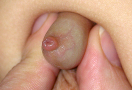
Uneven preputial separation may result in glanular adhesions between the glans and the prepuce, and a whitish collection of desquamated cells called smegma may be visible between the glans and the prepuce. The preputial skin is usually supple, and urine trapping with ballooning of the foreskin may occur with urination.
In contrast, in severe (pathological) phimosis the foreskin is characterized by a sclerotic, white ring (cicatrix) at the tip.[Figure caption and citation for the preceding image starts]: Pathological phimosis with cicatrixFrom the collection of Warren T. Snodgrass, MD [Citation ends].
Paraphimosis
Paraphimosis typically presents as a swollen distal glans with retracted foreskin behind the glans, forming a constricting band. The oedema can be severe enough to distort normal anatomy such that circumcised or non-circumcised status of the penis may be indiscernible.
Serious complications such as necrosis of skin may rarely occur. Penile necrosis may occur after prolonged untreated paraphimosis and should be suspected with a firm, discoloured glans.[41][Figure caption and citation for the preceding image starts]: ParaphimosisFrom the collection of Nicol Corbin Bush, MD [Citation ends].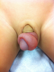 [Figure caption and citation for the preceding image starts]: Early paraphimosisFrom the collection of Nicol Corbin Bush, MD [Citation ends].
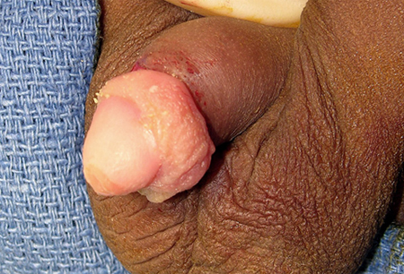
Hypospadias
Hypospadias is typically diagnosed at birth when the hallmark incomplete prepuce is visualised. Severity can range from the distal form (i.e., the urethral meatus is located on the glans or distal shaft) to the less common proximal form (i.e., the urethral meatus is located from the perineum to the proximal shaft).
Incomplete prepuce is a frequent finding with both proximal and distal hypospadias, though it may be intact in some cases of distal hypospadias. Penile curvature is also common, particularly in patients with proximal hypospadias; patients with proximal hypospadias may also have a cleft scrotum. Megameatus with intact prepuce (a mild variant of distal hypospadias) can occur with an intact prepuce, and is only noted at the time of circumcision or retraction of the prepuce at an older age.[Figure caption and citation for the preceding image starts]: Infant with distal hypospadias. The urethral meatus is located in the glans or distal shaft and prepuce is typically incompleteFrom the collection of Nicol Corbin Bush, MD [Citation ends].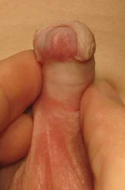 [Figure caption and citation for the preceding image starts]: Infant with proximal hypospadias. The urethral meatus is located from the perineum to the proximal shaftFrom the collection of Nicol Corbin Bush, MD [Citation ends].
Classification of hypospadias and its associated findings continues to evolve, with the Glans-Urethral Meatus-Shaft Score (GMS Score) and photographic analysis currently in use.[2][3][43][44]
Congenital penile curvature and/or torsion
Congenital penile curvature and/or torsion are typically noted in the newborn period when the infant has a spontaneous erection, often with voiding. A forward or sideways penile bend is seen, sometimes with penoscrotal webbing and/or deficient ventral shaft skin. Less commonly, dorsal curvature is seen. Penile curvature may occur in conjunction with hypospadias, or as an isolated finding. The position of the meatus should be evaluated in these patients.
Congenital penile curvature >30 degrees is considered clinically significant.[11][Figure caption and citation for the preceding image starts]: Congenital penile curvature (congenital chordee)From the collection of Nicol Corbin Bush, MD [Citation ends].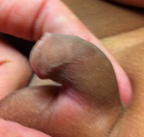
Concealed or buried penis
In pre-pubertal boys, overweight adolescents, and men (with class III obesity [BMI 40 or above]), the shaft and/or glans penis may be covered by an enlarged suprapubic fat pad in a condition known as buried penis; this can occur in both circumcised and uncircumcised males. Although the penis may not be visible, it can usually be fully inspected by retraction of the skin on either side of the penis by placing the thumb and forefinger on either side at the base of the penis and pushing proximally along the penile shaft to the level of the pubic symphysis. The penis can then usually be assessed either by inspection or by further palpation to assess the true length of the penile corpora, although this may be difficult in obese patients in whom pathological phimosis is also present.
Distortion of normal anatomy leading to secondary buried penis also occurs in the presence of hernia or hydrocele and usually resolves with repair of the hernia or hydrocele.
Congenital buried penis (also known as congenital megaprepuce) is characterised by excessive inner preputial skin associated with a tight phimotic ring in newborns, leading to urine entrapment appearing visually as severe ballooning of the penile skin and scrotum with urination. Urine trapped within the penile and scrotal skin may need to be manually expressed. This is a rare condition that requires surgical referral for correction.[Figure caption and citation for the preceding image starts]: Congenital buried penisFrom the collection of Nicol Corbin Bush, MD [Citation ends].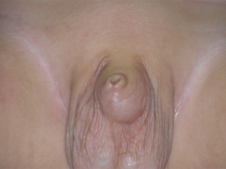 [Figure caption and citation for the preceding image starts]: Congenital buried penis: abundant inner preputial skin with paucity of shaft skinFrom the collection of Nicol Corbin Bush, MD [Citation ends].
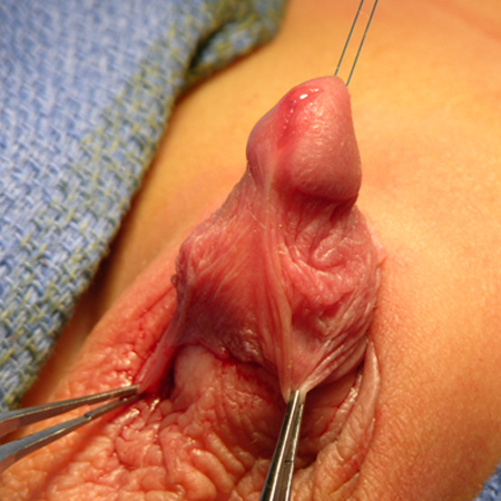 [Figure caption and citation for the preceding image starts]: Congenital buried penisFrom the collection of Warren T. Snodgrass, MD [Citation ends].
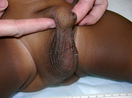 [Figure caption and citation for the preceding image starts]: Congenital buried penisFrom the collection of Nicol Corbin Bush, MD [Citation ends].
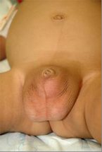 [Figure caption and citation for the preceding image starts]: Repaired congenital buried penis (with bilateral hernia repair and scrotoplasty)From the collection of Warren T. Snodgrass, MD [Citation ends].
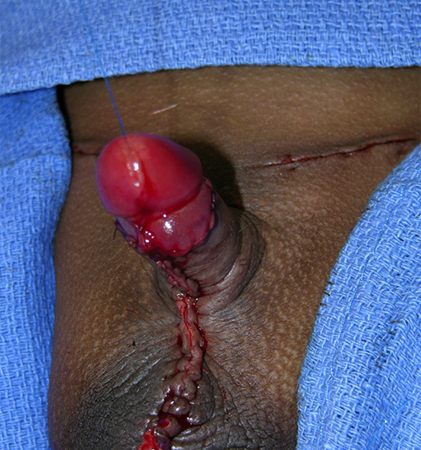 [Figure caption and citation for the preceding image starts]: Repaired congenital buried penisFrom the collection of Nicol Corbin Bush, MD [Citation ends].
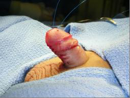
Micropenis
A buried penis and a micropenis can be distinguished by careful measurement of the penile shaft (not the penile skin).
If the stretched penile length is 2.5 or more standard deviations below the mean for age, micropenis is diagnosed and endocrine evaluation is appropriate.[45]
If the penis is of normal length but appears short because it is buried within surrounding tissues, the diagnosis of concealed penis is made.
Laboratory and tests
The diagnosis of anatomical penile abnormalities is based solely on history and physical examination.
Patients with hypospadias and unilateral cryptorchidism with a palpable testis can see a surgical specialist for further evaluation, and frequently benefit from endocrine evaluation. Infants with hypospadias and bilateral non-palpable gonads should undergo immediate endocrine evaluation.
Use of this content is subject to our disclaimer