Approach
Diagnosis is usually clinical based on the following presentation:[34]
Typical anatomical location (axillae and inguinal regions; symmetrical lesions suggest systemic disease rather than local infection)
Relapses and chronicity
Typical lesions (deep-seated nodules [boils], comedones, and scarring).
History
For all patients, a full medical and medication history should be taken. Recognised risk factors for HS (e.g., obesity, smoking, positive family history) should be noted.
Patients typically present with painful, firm nodules in the axilla, perineum, and/or groin. Women may report that tenderness and swelling begin about 1 week prior to the onset of menses (premenstrual flare).[35] Patients may experience discomfort or pruritus in the affected region prior to the nodules forming.
Many patients may report previous episodes that may have been unresponsive to antibiotic therapy or that required surgical drainage.[1] Some patients may also report treatment for acne vulgaris.
Comorbidity assessment
Prompt diagnosis and treatment of concomitant disease benefits patient health and quality of life. All patients should be screened for metabolic syndrome, type 2 diabetes, hypertension, Crohn's disease, and depression.[36][37]
Physical examination
Inflammatory nodules and abscesses may be present in the intertriginous areas (axilla, groin, perineum, or infra-mammary region). Prominent open comedones are often found. Non-intertriginous areas are rarely affected.
Although HS may be unilateral in early disease, most cases will progress to involve both sides of the body. Fibrosis, dermal contractures, disfiguring scars, and sinus tracts may be seen in long-standing disease.
In persistent peri-anal disease other diagnoses including Crohn's should be considered. [Figure caption and citation for the preceding image starts]: Hidradenitis suppurativa stage I: discrete inflamed nodules and papules with intervening normal skin and lack of scarringFrom R.A. Lee, MD, PhD [Citation ends]. [Figure caption and citation for the preceding image starts]: Hidradenitis suppurativa stage II: inflamed nodules and scars with areas of intervening normal skinFrom R.A. Lee, MD, PhD [Citation ends].
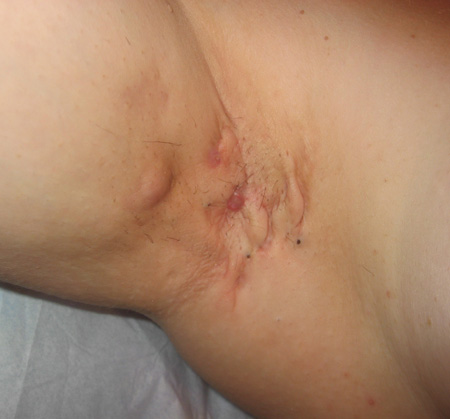 [Figure caption and citation for the preceding image starts]: Hidradenitis suppurativa: fluctuant abscess in axillaFrom R.A. Lee, MD, PhD [Citation ends].
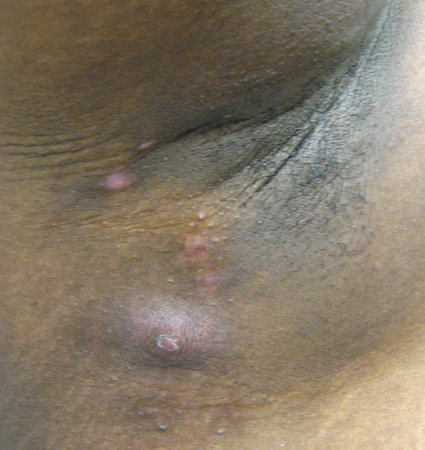 [Figure caption and citation for the preceding image starts]: Hidradenitis suppurativa: open 'double' comedonesFrom R.A. Lee, MD, PhD [Citation ends].
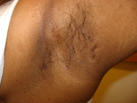 [Figure caption and citation for the preceding image starts]: Hidradenitis suppurativa: linear scarsFrom R.A. Lee, MD, PhD [Citation ends].
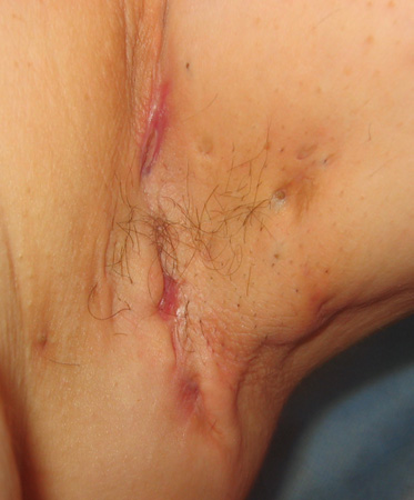 [Figure caption and citation for the preceding image starts]: Hidradenitis suppurativa: draining sinus tractsFrom R.A. Lee, MD, PhD [Citation ends].
Investigations
Targeted investigations may be indicated.
Bacterial cultures: can be useful to exclude MRSA furunculosis, and to document change in flora in response to long-term antibiotics.
Skin biopsy: may be performed in select patients to exclude squamous cell carcinoma (SCC) and to differentiate from other inflammatory diseases, including Crohn's disease. SCC may be a consequence of long-standing chronic inflammation in advanced HS.[38][39]
Low zinc levels can manifest as chronic skin erythema in intertriginous areas (acrodermatitis enteropathica); some data suggest that low serum zinc levels are more prevalent in HS than in a healthy population.[40]
Use of this content is subject to our disclaimer