Case history
Case history
A 65-year-old man presents with acute pain and swelling in the left leg for the past 3 days. The pain is localised in the posteromedial calf and worsens during standing and walking. There are no known risk factors for deep vein thrombosis (DVT), and no history of previous DVT or trauma. Past medical history is significant for hypertension and osteoarthritis of both knees. Medications include metoprolol and non-steroidal anti-inflammatory drugs (NSAIDs). Physical examination reveals a normal vascular examination, absent leg oedema, calf tenderness, and a prominent non-pulsatile mass behind the knee. Duplex ultrasound identifies a cyst in the popliteal crease measuring 4 cm x 5 cm.
Other presentations
Pseudothrombophlebitis syndrome is an acute presentation of pain, oedema, and erythema of the calf. This mimics DVT of the calf and can occur with rupture of a cyst or an extensive dissecting unruptured cyst. Imaging (ultrasound or magnetic resonance imaging) helps to distinguish between DVT and pseudothrombophlebitis syndrome. Pseudothrombophlebitis demonstrates a normal venous duplex examination and the dissecting fluid emanating from the ruptured cyst is usually visualised.
Large cysts may compress the popliteal vein, but this is very rare because the cyst develops medial to the popliteal vessels.[Figure caption and citation for the preceding image starts]: Anatomical dissection of the posteromedial knee capsule. The weak area (A) is identified between the two expansions of the semimembranosus muscle (B), the oblique popliteal ligament (C), and the expansion over the sheath of the popliteus muscle (D). The semitendinosus muscle (E) and popliteus muscle (F) are also indicatedAdapted from Labropoulos N, Shifrin DA, Paxinos O. New insights into the development of popliteal cysts. Br J Surg. 2004;91:1313-1318; used with permission [Citation ends].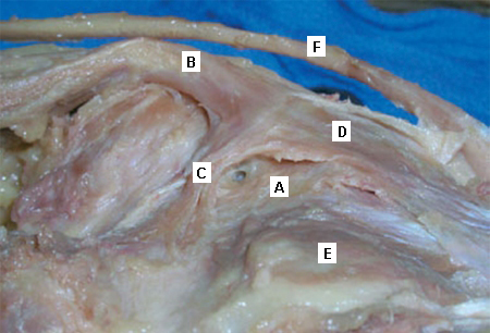 [Figure caption and citation for the preceding image starts]: A ruptured haemorrhagic popliteal cyst extending in the calfStony Brook University Medical Center private collection; used with permission [Citation ends].
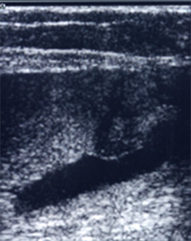
Use of this content is subject to our disclaimer