History and exam
Key diagnostic factors
common
presence of risk factors
White ethnicity, female sex, prematurity, low birth weight, multiple gestation, and advanced maternal age are strong risk factors for development of haemangioma.
variable onset
Haemangioma promontory marks/signs may be present at birth, but they more typically present during the first few weeks of life.[2][16] These initial clinical signs may be subtle pink patches or telangiectasias. They are also sometimes thought to be bruises or secondary to birth trauma. Deep haemangiomas appear later, in the first few months of life.
rapid growth
variable compressibility
An infantile haemangioma is soft and not fixed. Superficial haemangiomas develop a tight and tense surface with time, whereas deep haemangiomas feel tense and may swell with crying or dependency. With involution, the haemangioma is palpably softer.
flat or nodular character
Present as flat macules or patches; alternatively, the haemangioma may present as a papule or nodule.
Other diagnostic factors
common
islands of normal skin
ulceration and bleeding
Ulceration and bleeding, particularly in areas subject to increased friction and trauma.
warmth
Haemangiomas may develop into raised, warm, firm-textured lesions.
uncommon
history of low birth weight
Studies suggest that low birth weight is a significant risk factor.[14]
variable pain
associated defects
PHACES syndrome (posterior fossa malformations, haemangioma, cardiac defects and coarctation of the aorta, eye anomalies, sternal defects, and supra-umbilical raphe) has been associated with large, plaque-type cervicofacial haemangioma.[1][Figure caption and citation for the preceding image starts]: Tender, ulcerated haemangioma on the left lower lipFrom the collection of Carla T. Lane, MD, PhD; used with permission [Citation ends].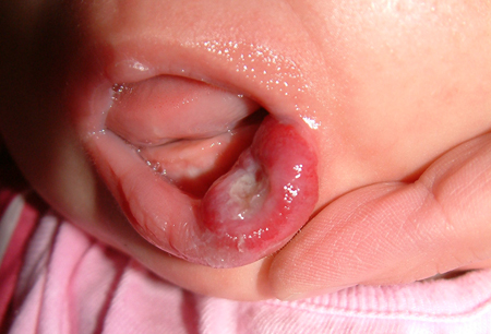
beard distribution and stridor
Haemangiomas located on the lower face and neck have been associated with laryngeal haemangioma.
lumbosacral location
Haemangioma located in the lumbosacral area may signal underlying spinal dysraphism. Other associated malformations include tethered cord, and renal and skeletal anomalies (LUMBAR syndrome).
multiple lesions
Infants who present with multiple haemangiomas should be screened for visceral lesions as part of a constellation called multifocal infantile haemangiomas with extracutaneous involvement.
poor infantile feeding, failure to gain weight
Multifocal infantile haemangiomas with extracutaneous involvement may present with gastrointestinal involvement. Oral and lip lesions may cause difficulty feeding.
high-output cardiac failure
Unrecognised, large, proliferative visceral haemangioma may lead to high-output cardiac failure.
Risk factors
strong
low birth weight
white ethnicity
female sex
maternal multiple gestation
weak
chorionic villus sampling
Though traditional thinking has held that chorionic villus sampling is contributory, more recent research has undermined the significance of chorionic villus sampling during pregnancy as an associated risk factor for haemangioma.[17]
Use of this content is subject to our disclaimer