Aetiology
The aetiology of haemangioma is poorly understood. Both intrinsic and extrinsic mechanisms have been proposed. There is much debate regarding the origin of the precursor cell to haemangioma.
Pathophysiology
Infantile haemangioma may originate from the embryonic mesoderm, a pericyte or endothelial precursor, or localised angioblasts.
According to the intrinsic theory, infantile haemangioma originates from vasculogenesis, a process by which new blood vessels are formed.
Extrinsic theory suggests that external environmental factors provide an environment favourable for the development of infantile haemangioma. Proposed stimulants include hypoxia, local growth factors, cytokines, and oestrogens. Haemangioma could develop as a result of vasculogenesis or angiogenesis, whereby new blood vessels arise from existing ones.
Within both intrinsic and extrinsic theories, the precursor cell of the infantile haemangioma originates from the embryo.[14][20] Several characteristic cellular placental markers have been identified in infantile haemangioma. Debate exists over whether the precursor cell may be a displaced placental angioblast; however, molecular genetic studies of infantile haemangioma point towards an embryonic origin for the precursor cell. Haemangioma endothelial cells (HECs) from male infants contain XY chromosomes. Genetic polymorphisms of the HECs match those of the child, not the mother. Thus, if the precursor cell arises from the embryo, it may develop towards a placental phenotype.[21][22]
Classification
Infantile haemangiomas are classified as vascular tumours in the International Society for the Study of Vascular Anomalies classification, which was last updated in April 2014.
International Society for the Study of Vascular Anomalies: classification for vascular anomalies
Opens in new window Tumours in this classification structure share the characteristics of growth and endothelial proliferation.[Figure caption and citation for the preceding image starts]: Examples of infantile haemangiomas according to International Society for the Study of Vascular Anomalies classificationInternational Society for the Study of Vascular Anomalies; used with permission [Citation ends].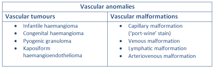
Clinical classification[4][5]
Superficial
Most common type of haemangioma, comprising 43% of total; located in the superficial dermis and characterised by bright red colour in the growth phase.
Deep
16% of total, located in the deep dermis and subcutaneous tissues; may present later in life as blue nodule.
Mixed (superficial and deep)
41% of total; appear as combination of deep blue nodules with overlying bright red superficial plaque.
Morphological classification[6][7]
Localised
Arises from a single focus; often round or oval in shape.
Segmental
Seem to arise from a broad area; can be associated with underlying abnormalities such as those seen in PHACES syndrome (posterior fossa malformations, haemangioma, arterial anomalies, coarctation of the aorta and cardiac defects, eye abnormalities, and sternoclavicular or supraumbilical anomalies) or LUMBAR syndrome (lower body infantile haemangioma and other cutaneous defects, urogenital anomalies and ulceration, myelopathy, bony deformities, anorectal malformations and arterial anomalies, and renal anomalies).[1]
Indeterminate
Ambiguity in characteristics that defies classification as focal or segmental.
Clinical variants and special considerations
Segmental haemangiomas may have associated underlying abnormalities. Segmental cervicofacial haemangiomas may have associated structural anomalies of the brain, cerebral vasculature, eyes, sternum, and/or aorta. This neurocutaneous disorder is known as PHACE(S) syndrome, with the acronym referring to posterior fossa anomalies, haemangioma, arterial lesions, cardiac abnormalities/aortic coarctation, abnormalities of the eye, and sternal clefting or supraumbilical raphe.[8] Infants with segmental cervicofacial haemangioma require ophthalmological examination, echocardiogram, and possible central nervous system imaging.
Beard haemangioma: haemangiomas located on the lower face and neck have been associated with laryngeal haemangioma.[1] Progressive stridor is a worrisome sign. Infants with haemangiomas in beard distribution should be referred to an otolaryngologist for further evaluation and possible endoscopy.[1]
Lumbosacral haemangioma: haemangioma located in the lumbosacral area may signal underlying spinal dysraphism. Other associated malformations include tethered cord, renal, and skeletal anomalies. Magnetic resonance imaging is the test of choice.[1][9][10] Segmental perineal haemangiomas should raise concern for LUMBAR syndrome, which refers to lower body infantile haemangioma and other cutaneous defects, urogenital anomalies and ulceration, myelopathy, bony deformities, anorectal malformations and arterial anomalies, and renal anomalies.[1]
Multifocal infantile haemangiomas (previously termed diffuse neonatal haemangiomatosis): Infants with multiple cutaneous haemangiomas may have haemangiomas within their visceral organs.[11] A prospective study revealed that 16% of infants who present with ≥5 infantile haemangiomas have hepatic haemangiomas.[12] In such patients a good physical examination is indicated. Hepatomegaly may indicate clinically significant liver haemangiomas and should be evaluated by ultrasound. An abnormal cardiac examination may indicate high-output heart failure.
Haemangiomas in certain locations can result in significant cosmetic or functional complications. Periorbital haemangiomas may result in ocular complications. Haemangiomas on the nasal tip or ear may cause cartilage destruction and permanent disfigurement. Lip haemangiomas can distort the normal contour of the mouth.[13] Genital and perineal haemangiomas are more likely to ulcerate and lead to associated complications. [Figure caption and citation for the preceding image starts]: Plaque-type cervicofacial ulcerated haemangioma (beard distribution)From the collection of Carla T. Lane, MD, PhD; used with permission [Citation ends].
Use of this content is subject to our disclaimer