Aetiology
A decreased salivary flow rate can be caused by dehydration, malnutrition, and immunosuppression. A number of therapeutic drugs (e.g., diuretics, antihistamines, antidepressants, and antihypertensives) may result in pharmacologically induced salivary gland hypofunction.[20] Decrease of salivary output can facilitate the ascending retrograde bacterial colonisation of the salivary gland parenchyma through the ductal system.
Mechanical obstruction due to sialolithiasis or ductal anomalies (e.g., sialectasis, diverticuli, and strictures) may also reduce the salivary output, and thus predispose the individual to an ascending bacterial sialadenitis.[21][20] Acute adult suppurative sialadenitis may be caused by aerobic or facultative bacteria, anaerobic bacteria, or both. Typical aerobic micro-organisms are Staphylococcus aureus and Haemophilus influenzae. Gram-negative bacilli include pigmented Prevotella, Porphyromonas, and Fusobacterium.[22] In neonates, Staphylococcus aureus and occasionally Pseudomonas aeruginosa are often the cause.[10][12] In children, the parotid gland is most frequently affected by acute bacterial infection, typically with Staphylococcus aureus or group A streptococci.[23]
Surgical interventions in debilitated people are one of the most common predisposing factors for the development of acute sialadenitis in a hospital setting; this is potentially due to a number of factors, such as lack of salivary stimulation due to the patient not eating.[24][25] General anaesthesia can predispose to the development of acute sialadenitis. Acute sialadenitis has also been reported after cheek biting, accidental introduction of air into major salivary glands during dental procedures, and orthodontic movement.[20]
Chronic recurrent episodes of acute inflammation may result from underlying ductal abnormalities or may be associated with Sjogren syndrome. Sjogren syndrome is more common in peri- and post-menopausal women.[3]
IgG4-related disease has been recognised as a rare cause of persistent bilateral, painless salivary gland swelling in middle aged or older males. There may be single or multi-organ involvement with the lacrimal and periorbital tissues often involved in the head and neck region.[6] Chronic sclerosing sialadenitis (Kuttner's tumour) is a localised form of IgG4-related sclerosing disease.[7]
The aetiology of autoimmune sialadenitis is unknown but may be associated with xerostomia, xerophthalmia, and connective tissue diseases (e.g., SLE, rheumatoid arthritis, and scleroderma). Intraductal nidus of debris composed of inspissated mucus, bacteria, ductal epithelial cells, or foreign bodies may become calcified and form sialoliths (ductal stones). Calcium and phosphorus metabolism is normal in these patients.[26] The aetiology of subacute necrotising sialadenitis (SANS) is unknown, but infectious and allergic aetiologies have been suggested. SANS is an unusual self-limiting inflammatory condition seen commonly in teenagers and young adults. It is considered separate entity to necrotising sialadenitis.[27]
Pathophysiology
The initial stage of acute bacterial sialadenitis is characterised by accumulation of bacteria, neutrophils, and inspissated fluid in the lumen of ductal structures. Ductal epithelium damage gives rise to sialodochitis (periductal inflammation), accumulation of neutrophils in the glandular stroma, and subsequent acini (secretory units of salivary glands) necrosis with formation of microabscesses.[13] The chronic stage is established with recurrent episodes and is characterised by further destruction of salivary acini and the establishment of periductal lymph follicles.[13] In chronic sclerosing sialadenitis, various degrees of inflammation (ranging from focal lymphocytic sialadenitis to widespread salivary gland cirrhosis with effacement of acini) can result from obstruction of the salivary ducts by microliths, from associated intercurrent infections, or from immune reaction with the formation of secondary lymph follicles.[13] In autoimmune sialadenitis, a response to an unidentified antigen present in the salivary gland parenchyma results in activation of T and B cells that infiltrate the interstitium, with ensuing acini destruction and the formation of epimyoepithelial islands. This increases the likelihood of developing B-cell lymphoma.[13]
Classification
Aetiological and histological classification of sialadenitis[1]
Bacterial sialadenitis
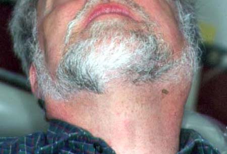
Divided into acute and chronic subtypes. Acute bacterial sialadenitis has a predilection for the parotid glands of children and older adults, with two distinct presentations: nosocomial and community-acquired.[Figure caption and citation for the preceding image starts]: Acute bacterial sialadenitis of left parotid glandFrom the personal collection of Dr A. Aguirre; used with permission [Citation ends].
 Chronic infection may result in recurrent sialadenitis as the ongoing low-grade infection can cause sporadic clinically detectable episodes.
Chronic recurrent sialadenitis
Mainly presents in adults (only 10% of patients are children). It is typically tender, unilateral swelling of a major salivary gland of an episodic nature. It represents recurrent episodes of acute sialadenitis. This may be due to unresolved infection or underlying ductal anomalies.
Obstructive sialadenitis
Obstructive sialadenitis caused by stones has a predilection for submandibular and parotid glands. Typically, it is a unilateral painful enlargement occurring in connection with eating.[2]
Classical autoimmune sialadenitis
Classical autoimmune sialadenitis such as Sjogren syndrome typically affects the salivary and lacrimal glands and primarily occurs in adult women and is linked to peri- and post-menopause.[3]
Sjogren syndrome may be primary or secondary in association with another autoimmune disease, such as systemic lupus erythematosus, rheumatoid arthritis, or scleroderma.[4] Data suggests that approximately 25% of patients with rheumatoid arthritis or systemic lupus erythematosus have histological evidence of Sjogren syndrome.[5]
In contrast, IgG4-related disease is a relatively newly described fibro-inflammatory disorder that typically affects middle-aged males and is characterised by unilateral or bilateral persistent painless swellings of the parotid and/or submandibular glands.[6]
Chronic sclerosing sialadenitis (Kuttner's tumour)
A localised form of IgG4-related sclerosing disease. It has a predilection for submandibular glands.[7] Typically, it is a unilateral enlargement that may be symptomatic and clinically difficult to differentiate from a tumour.
Subacute necrotising sialadenitis
Subacute necrotising sialadenitis is a rare condition affecting the palatal salivary glands. May be an early form of necrotising sialometaplasia. Presents as a lump on the hard or soft palate, usually painful but only occasionally ulcerated. Aetiology is unknown and it resolves within a few weeks.[8]
Use of this content is subject to our disclaimer