Images and videos
Images
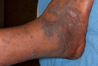


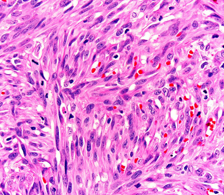
Kaposi's sarcoma
Photomicrograph of the histopathology of Kaposi's sarcoma showing fascicles of vasoformative spindle-shaped tumour cells (haematoxylin and eosin stain)
From the collection of Dr Liron Pantanowitz; used with permission
See this image in context in the following section/s:

Kaposi's sarcoma
Kaposi's sarcoma cutaneous purple-brown plaque on the foot
From the collection of Dr Bruce J. Dezube; used with permission
See this image in context in the following section/s:
Kaposi's sarcoma
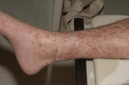
Multiple pink-purple Kaposi's sarcoma nodules on the lower extremity
From the collection of Dr Bruce J. Dezube; used with permission
See this image in context in the following section/s:
Kaposi's sarcoma
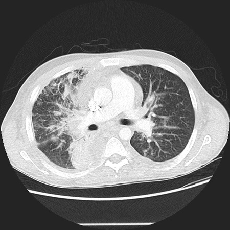
CT scan of the chest showing a reticular pattern due to pulmonary involvement by Kaposi's sarcoma
From the collection of Dr Bruce J. Dezube; used with permission
See this image in context in the following section/s:
Use of this content is subject to our disclaimer