Investigations
1st investigations to order
CXR
Test
Chest radiography is the first-line imaging modality in any patient presenting with known trauma.[43]
It helps detect the rib fracture, but conventional chest x-rays can miss up to 50% of rib fractures.[44]
It is not usually necessary to perform dedicated rib radiography, in addition to chest radiography, for the diagnosis of rib fractures in adults after minor trauma.[43]
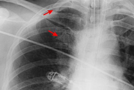
[Figure caption and citation for the preceding image starts]: CXR showing right first rib fractureFrom the collection of Dr Paul Novakovich; used with permission [Citation ends].
 [Figure caption and citation for the preceding image starts]: CXR showing multiple posterior left-sided rib fracturesFrom the collection of Dr Paul Novakovich; used with permission [Citation ends].
 [Figure caption and citation for the preceding image starts]: Anteroposterior CXR multiple left-sided rib fractures with chest tube in placeFrom the collection of Dr Paul Novakovich; used with permission [Citation ends].
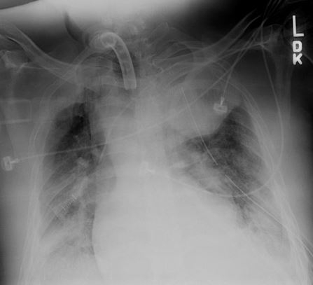
Also assess for pneumothorax, haemothorax, and aortic injury.
Practical tip
In practice, a single rib fracture with a clear history, such as following a fall, might be made as a clinical diagnosis, even if no fracture is identified on a chest x-ray.
Result
rib fracture ± associated parenchymal injury; pneumothorax; effusion/haemothorax
CT chest
Test
Use an early CT scan with contrast in adults presenting with blunt chest wall trauma and for adults with suspected chest trauma without severe respiratory compromise who are responding to resuscitation or whose haemodynamic status is normal.[33] In practice, most patients can be stabilised sufficiently in order to receive a CT scan.
CT scan of the chest can improve the sensitivity in detecting rib fractures as well as other injuries.
Consider it if there are clinical features suggestive of rib fracture and there is the potential for improved patient care if rib fracture(s) are detected.
CT imparts significant radiation exposure to the patient and in a patient with minor trauma the increased sensitivity of CT does not necessarily alter management or clinical outcomes of patients who do not have associated injuries.[43]
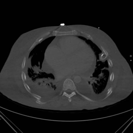
[Figure caption and citation for the preceding image starts]: CT scan showing bilateral posterior rib fracturesFrom the collection of Dr Paul Novakovich; used with permission [Citation ends].
 [Figure caption and citation for the preceding image starts]: CT showing right anterolateral rib fractureFrom the collection of Dr Paul Novakovich; used with permission [Citation ends].
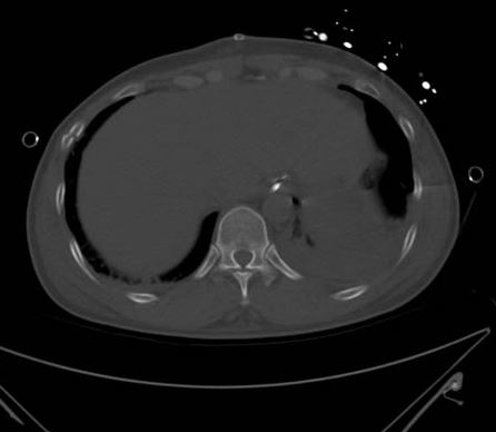 [Figure caption and citation for the preceding image starts]: CT showing left posterior segmental rib fractureFrom the collection of Dr Paul Novakovich; used with permission [Citation ends].
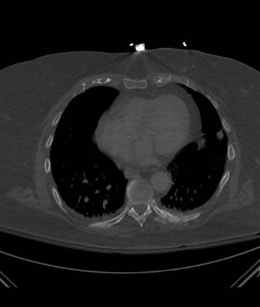 [Figure caption and citation for the preceding image starts]: CT scan showing large left-sided pneumothoraxFrom the collection of Dr Paul Novakovich; used with permission [Citation ends].
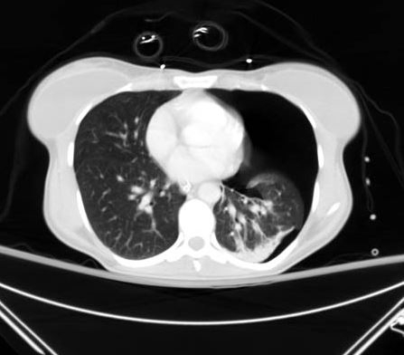 [Figure caption and citation for the preceding image starts]: CXR depicting the same pneumothorax as shown on CTFrom the collection of Dr Paul Novakovich; used with permission [Citation ends].
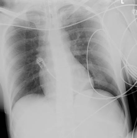
In a stable patient who has sustained a high-energy impact trauma, request a CT scan of the head, cervical spine, chest, abdomen, and pelvis to be performed in adults to rule out other injuries.
Do not routinely use CT for first‑line imaging to assess chest trauma in children.[33]
Result
fracture ± associated parenchymal injury
extended focused assessment with sonography
Test
Consider immediate extended focused assessment with sonography for trauma (eFAST) to assess chest trauma in adults with severe respiratory compromise.[33] In practice, the role of point-of-care ultrasonography may be in the immediate assessment of a trauma patient; it is quick and easily performed at the bedside, and can be performed alongside resuscitation.[42] However, it is operator dependent, and more research is needed to confirm its accuracy in diagnosing thoracic trauma.[42]
Result
may identify pneumothorax, haemothorax, or bleeding
Investigations to consider
CT of head, cervical spine, abdomen, and pelvis
Test
If the patient is stable, a CT scan of the head, cervical spine, chest, abdomen, and pelvis should be performed in adults presenting after high-energy impact to rule out other injury.
Result
may reveal other injuries
echocardiography
Test
Consider echocardiography in patients with a clinical or radiological suspicion of cardiac injury.
Result
may reveal cardiac contusion, pericardial tamponade, or acute valvular injury, as a consequence of the fracture
skeletal survey and CT head (children)
Test
Any rib fracture in a child or infant should be assumed to be the result of non-accidental injury until shown otherwise.
Children with suspected maltreatment or non-accidental injury should be investigated according to local safeguarding policies. This would include a skeletal survey in all children aged under 2 years (and on a case-by-case basis in older children) and a CT head scan in children aged under 1 year or if there is external evidence of head trauma and/or abnormal neurological symptoms or signs.[45] This imaging should be requested by a senior clinician (usually a paediatrician).[45]
Result
may reveal other injuries
Use of this content is subject to our disclaimer