Etiology
Human African trypanosomiasis is caused by protozoa belonging to the genus Trypanosoma, species brucei. There are two human pathogen subspecies: T b gambiense and T b rhodesiense. They are extracellular parasites. Traditionally, only the rhodesiense and gambiense subspecies cause sleeping sickness. Human infections with the closely related animal trypanosomes T b brucei, T evansi, and T congolense have been reported, but are rare. Infections are probably associated with the absence or decrease of apolipoprotein L-1, which has been shown to be the trypanolytic factor in human blood, but they have also been observed in immunosuppressed adults and children.[8][9][10]
The disease is transmitted cyclically, by the bite of a tsetse fly (genus Glossina). Transmission is mainly anthroponotic for gambiense trypanosomiasis, in which the animal reservoir plays a minor role, and zoonotic for the rhodesiense form, for which wild animals and cattle are the main reservoir.[Figure caption and citation for the preceding image starts]: Tsetse fly (Glossina)Published with permission from M. Madder PhD, Institute of Tropical Medicine, Antwerp, Belgium [Citation ends].
Other modes of transmission such as mother to child, blood transfusion, or laboratory infection are rare. Mechanical transmission by other insects and sexual transmission have been described but are controversial.[11][12][13]
Pathophysiology
When tsetse flies bite an infected mammalian host, they ingest trypanosomes with the host's blood. After several differentiation steps in the fly, trypanosomes reach the salivary glands. With every subsequent bite, the infected tsetse fly injects trypanosomes with the saliva.[14][Figure caption and citation for the preceding image starts]: Life cycle of T b gambiense and T b rhodesiensePublished with permission from the CDC [Citation ends].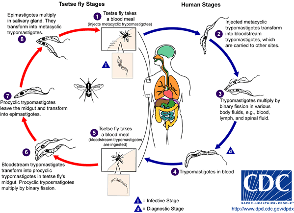
At the inoculation site, because of the localized inflammatory reaction, a local chancre may form, which spontaneously resolves within days to weeks. Trypanosomes multiply and spread to the lymphatic system and bloodstream. In the bloodstream, the surface of trypanosomes is entirely covered with variant surface glycoprotein (VSG), which forms a coat of identical proteins. The host mounts an effective antibody response to the VSG, resulting in opsonization (marking of the antigen for destruction by phagocytes) and parasite clearance. Trypanosomes are capable of evading this immune response by antigenic variation: they spontaneously switch their VSG coat, which is then no longer recognized by existing antibodies.[15][16] The interplay between the host's antibody response and the parasite's antigenic variation results in peaks of parasitemia. As well as antibodies against VSG, the polyclonal B-cell activation gives rise to antibodies against invariant trypanosome antigens, but also to autoantibodies.
Progressively, the disease advances into an immunosuppression status with a hypergammaglobulinemia. The lymph nodes, liver, and spleen become enlarged, related to the immune activation. High concentrations of inflammatory (interferon-gamma and tumor necrosis factor [TNF]-alpha) and counterinflammatory cytokines (interleukin [IL]-10) appear. Hormonal changes are frequent and related to infiltration of endocrine organs (mainly thyroid and adrenals), and lately related to hypothalamic-hypophysial tract lesions and to alteration of circadian rhythm.
Eventually, the trypanosomes cross the blood-brain barrier and enter the central nervous system (CNS). They cause extensive meningoencephalitis, infiltration of white matter with inflammatory cells, perivascular cuffing, and astrocyte and macrophage stimulation. The site of invasion and the inflammatory reaction determine the appearance and type of neuropsychiatric symptoms. Brain invasion is accompanied by a strong intrathecal immunoglobulin response.[17][18] Production of cytokines (IL-10, IL-6) and chemokines (CXCL-8, CXCL-10, CXCL-13) in the CNS seems to be related to progression of the disease and severity of the neurologic signs.[19][20][21][22] If untreated, patients usually progress to coma, severe organ failure, and eventually death.[23][24]
Use of this content is subject to our disclaimer