Etiology
There is no specific identifiable causative factor; however, there is much speculation regarding etiology.
Traumatic events may be recalled and linked to the ganglion in anywhere from 10% to 40% of patients.[7]
Scapholunate injury has also been hypothesized to lead to dorsal ganglia.[8]
Arthrographic studies have demonstrated radiopaque dye passing from the wrist joint into the ganglion cyst unidirectionally, leading to the theory that a small hole in the wrist capsule may cause a one-way valve allowing growth of the cyst.[9]
Ganglions may also represent benign tumors of synovial origin; however, there is no specific synovial lining noted on histologic examination.[10]
Synovial fluid leaking into the surrounding tissue may lead to formation of a cyst wall enclosing cystic fluid.[11]
Mucoid degeneration of collagen in the surrounding tissue may lead to cyst formation.
A definitive link between ganglion cyst development and traumatic injury has not been established.
Pathophysiology
Ganglion cysts are usually small structures measuring 1 to 3 cm in diameter and located on the radial aspect. Impingement or surrounding of the radial artery can occur during cyst development. Occasionally, occult ganglions (<1 cm) or larger ganglions (up to 8 cm) have been reported. Cysts can be singular or multiloculated and are usually located next to a joint or the surrounding tendons. Macroscopically, they tend to be smooth, white and firm with an underlying stalk connection to the joint surface. They are mobile, nonadherent to underlying tissue, compressible, and usually not directly painful on palpation. The outer wall is comprised of randomly oriented collagen fibers with no definitive endothelial lining.[10] The cyst itself is filled with a thick, gelatinous, clear mucin comprised of glucosamine, globulin, hyaluronic acid, and albumin.[12]
Cysts may compress surrounding neurovascular structures and patients may experience wrist pain, paresthesia, intrinsic muscle paralysis, and/or coolness of the hand or fingers as a result.
Dorsally located cysts are usually connected to the scapholunate interosseous ligament in the area of the dorsal capsular attachments; however, the stalk may be long enough to have the palpable mass located a distance away from the dorsoradial wrist. Two thirds of volar cysts are connected to the radioscaphoid joint and one third to the scaphotrapezial joint.[13]
There is no documented history of malignant transformation of a ganglion cyst.
Classification
Anatomic
Volar: between the flexor carpi radialis tendon and the radial artery.
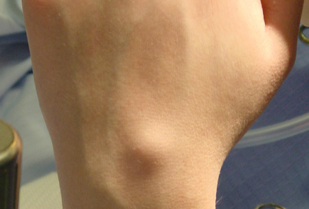
Dorsal: usually connected to the scapholunate ligament or scaphotrapeziotrapezoidal joint. [Figure caption and citation for the preceding image starts]: Typical dorsal wrist ganglion cystFrom the collection of Marco Rizzo, MD, Mayo Clinic; used with permission [Citation ends].
Occult: vague dorsal wrist pain with wrist flexion.
Use of this content is subject to our disclaimer